软骨组织由软骨细胞和软骨基质组成,软骨组织及其周围的软骨膜构成软骨,软骨染色的方法有很多种,如甲苯胺蓝法、阿利新蓝法、番红O法等。软骨根据基质内所含纤维素成分不同可分为透明软骨、弹性软骨、纤维软骨。透明软骨主要分布于上呼吸管道、肋骨内侧端、短骨关节面等处。
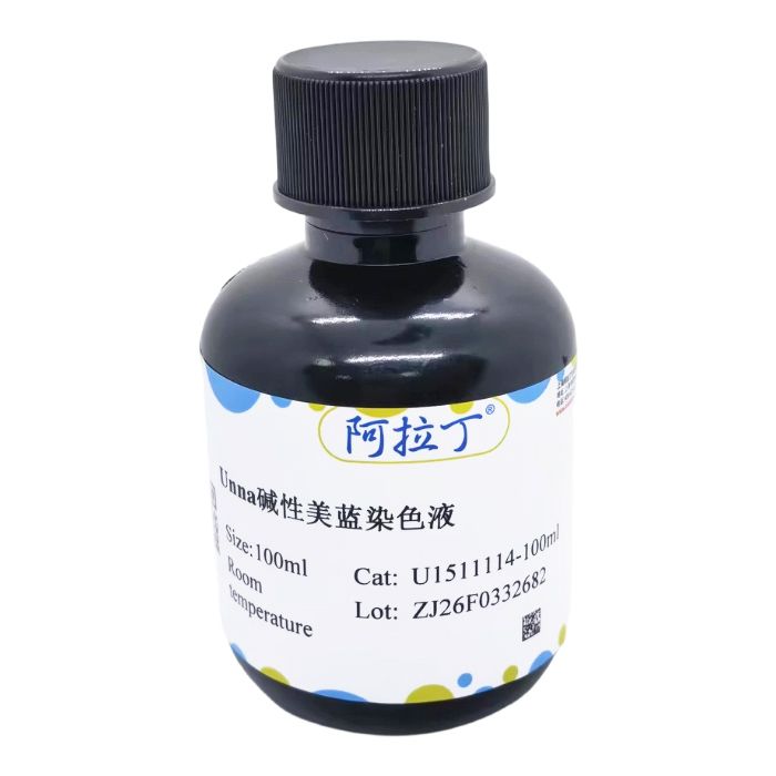
Unna碱性美蓝又称多色亚甲蓝 (Polychrome Methylene Blue),是亚甲基蓝的氧化混合物,通过特定工艺制备后呈现蓝、紫、红等多色性,在微生物学、病理学及材料科学中有独特应用。Unna碱性美蓝染色液可搭配茜素红用于透明软骨的染色,骨质(钙盐沉着处)和茜素红结合形成红色,软骨与多色亚甲蓝反应呈紫色,细胞质呈黄色,胞核蓝色,此法为6-8个月胎儿指骨最佳染色法。该试剂仅用于科研领域,不适用于临床诊断或其他用途。由碳酸钾、美蓝等组成。
自备材料:
1、系列乙醇、蒸馏水、二甲苯或环保脱蜡液、环保透明液、中性树胶、茜素红S染色液
2、显微镜
操作步骤(仅供参考):
1、组织用90%乙醇固定12~24小时,石蜡包埋切片。切片厚度3~5μm,脱蜡至水,蒸馏水洗。
2、入茜素红S染色液染色5~20min,蒸馏水稍洗。
3、入Unna碱性美蓝染色液复染2~6min,95%乙醇分色至染色清晰。
4、常规脱水,二甲苯或环保透明液,中性树胶封固。
染色结果:
钙盐沉着处呈红色,软骨基质呈紫色,细胞质呈黄色,细胞核蓝色。
注意事项:
1. 在不溶性钙染色中茜素红S的染色时间要根据钙盐的含量来确定,应在显微镜下观察,直到着色反应强烈而不弥散为止。
2、经过茜素红S染色液染色后,钙沉积物是双折射的。
3、Unna碱性美蓝染色液易挥发,染色时应加盖。
4、为了您的安全和健康,请穿实验服并戴一次性手套操作。
Cartilage tissue is composed of chondrocytes and cartilage matrix; cartilage tissue and the surrounding perichondrium together form cartilage. There are numerous staining methods for cartilage, such as the toluidine blue method, alcian blue method, safranin O method and others. Based on the differences in fibrous components contained in the matrix, cartilage is classified into hyaline cartilage, elastic cartilage and fibrocartilage. Hyaline cartilage is mainly distributed in the upper respiratory tract, the medial ends of ribs, the articular surfaces of short bones and other locations.
Unna Alkaline Methylene Blue Staining Solution, also known as Polychrome Methylene Blue, is an oxidized mixture of methylene blue. It exhibits polychromasia (blue, purple, red and other hues) after preparation via specific processes and has unique applications in microbiology, pathology and materials science. Unna's alkaline methylene blue staining solution can be used in combination with alizarin red for the staining of hyaline cartilage. Bone tissue (sites of calcium salt deposition) combines with alizarin red to appear red, cartilage reacts with polychrome methylene blue to appear purple, cytoplasm appears yellow and cell nuclei appear blue. This method is the optimal staining method for phalanges of 6–8 month-old fetuses. This reagent is for research use only and not intended for clinical diagnosis or any other applications. Composed of potassium carbonate, methylene blue and other components.
Materials to Be Prepared by the User:
1. Graded ethanol, distilled water, xylene or environment-friendly deparaffinization solution, environment-friendly clearing solution, neutral balsam, alizarin red S staining solution
2. Microscope
Operating Procedures (For Reference Only):
1. Fix the tissue in 90% ethanol for 12–24 hours, then perform paraffin embedding and sectioning. Cut sections at a thickness of 3–5 μm, deparaffinize to water and rinse with distilled water.
2. Immerse in alizarin red S staining solution for 5–20 minutes, then rinse briefly with distilled water.
3. Counterstain in Unna's alkaline methylene blue staining solution for 2–6 minutes, then differentiate with 95% ethanol until the staining is distinct.
4. Perform routine dehydration, clear with xylene or environment-friendly clearing solution, and mount with neutral balsam.
Staining Results:
Sites of calcium salt deposition appear red, cartilage matrix appears purple, cytoplasm appears yellow and cell nuclei appear blue.
Precautions:
1. For insoluble calcium staining, the staining time of alizarin red S should be determined according to the content of calcium salts. Observation under a microscope is required until the staining reaction is intense without diffusion.
2. After staining with alizarin red S staining solution, calcium deposits are birefringent.
3. Unna's alkaline methylene blue staining solution is volatile; a cover should be placed over the staining vessel during the staining process.
4. For your safety and health, wear a lab coat and disposable gloves during operation.

 ZJ26F0332682

 危险品化学品经营许可证(带存储)