Goat Anti-Rabbit IgG H&L Antibody | Goat Anti Rabbit IgG H&L Antibody | Anti-Rabbit IgG H&L Antibody | Anti Rabbit IgG H&L Antibody | Goat Anti-Rabbit IgG Antibody | Goat Anti Rabbit IgG Antibody | Anti- Rabbit IgG Antibody | Anti Rabbit IgG Antibody | AF
规格或纯度
ExactAb™, 已验证, Ex:401nm, Em:422nm, 1.0 mg/mL
生化机理
IgG 是血清和淋巴中含量最高的一类抗体。它能有效对抗细菌、真菌、病毒和外来颗粒。免疫球蛋白 G 抗体会触发补体系统的作用。
宿主种属
山羊(Goat)
种属反应性
兔(Rabbit)
免疫原
Rabbit IgG
偶联
AF405
Excitation(nm)
Violet Laser(405nm)
Emission(nm)
422nm
Ex/Em(nm)
Ex:401nm;Em:422nm
克隆类型
多克隆抗体
Format
Whole IgG
亚型
Goat IgG
纯化方法
antigen affinity chromatography
浓度
1.0 mg/mL
分子类型
抗体
产品线
生物产品
储存与运输
储存缓冲液
Liquid in PBS pH 7.4, containing 50% glycerol, 1% BSA and 0.05% Proclin300.
防腐剂
0.05% Proclin300
储存条件
避光,-20°C储存,避免反复冻融
运输条件
超低温运输
稳定性与储存
储存温度为 -20°C。避免冷冻/解冻循环。避免光照。
图片
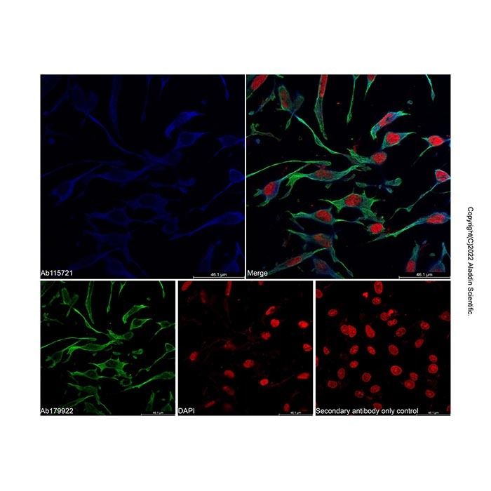
Goat Anti-Rabbit IgG H&L (AF405) (Ab181953) - IF IF analysis of MMP9 (blue) in U2OS cells. The cells were fixed and permeabilized with 100% methanol for 5 minutes, and blocked with 2% BSA for 1 hour at room temperature. Cells were stained with MMP9 antibody (Ab115721) at 1/500 dilution in blocking buffer overnight at 4°C, and then incubated with Goat Anti-Rabbit IgG H&L (AF405) (Ab181953) at a dilution of 1/1000 for 1 hour at room temperature in the dark. beta Tubulin Mouse mAb (Ab179922) (green) was stained with Goat Anti-Mouse IgG H&L (FITC) (Ab179003) at 1/1000 dilution. Cells were counterstained with PI (red). PBS instead of the primary antibody was used as the secondary antibody only control. Images were taken on the confocal laser scanning microscope.
Goat Anti-Rabbit IgG H&L (AF405) (Ab181953) - IF IF analysis of MMP9 (blue) in U2OS cells. The cells were fixed and permeabilized with 100% methanol for 5 minutes, and blocked with 2% BSA for 1 hour at room temperature. Cells were stained with MMP9 antibody (Ab115721) at 1/500 dilution in blocking buffer overnight at 4°C, and then incubated with Goat Anti-Rabbit IgG H&L (AF405) (Ab181953) at a dilution of 1/1000 for 1 hour at room temperature in the dark. Cells were counterstained with PI (red). PBS instead of the primary antibody was used as the secondary antibody only control. Images were taken on the confocal laser scanning microscope.


 ZJ24F0910381


 危险品化学品经营许可证(带存储)