Involved in the activation cascade of caspases responsible for apoptosis execution. Binding of caspase-9 to Apaf-1 leads to activation of the protease which then cleaves and activates effector caspases caspase-3 (CASP3) or caspase-7 (CASP7). Promotes DNA damage-induced apoptosis in a ABL1/c-Abl-dependent manner. Proteolytically cleaves poly(ADP-ribose) polymerase (PARP). Cleaves BIRC6 following inhibition of BIRC6-caspase binding by DIABLO/SMAC.
宿主种属
小鼠(Mouse)
特异性
Human Caspase-9
种属反应性
人(Human)
免疫原
Recombinant protein corresponding to Human Caspase-9
阳性对照
WB: HeLa, HepG2, K562 and HEK293 whole cell lysates Flow Cytometry: K562 cells IF/ICC: HeLa cells
偶联
Unconjugated
克隆类型
单克隆抗体
Format
Whole IgG
轻链亚型
kappa
纯化方法
Protein A purified
浓度
1.0 mg/mL
分子类型
抗体
产品线
生物产品
储存与运输
储存缓冲液
PBS, pH 7.2
储存条件
-20°C储存,避免反复冻融
运输条件
超低温运输
稳定性与储存
短期储存2-8℃ (1-2周); 长期储存-20℃ (24个月); 收货后建议分装,避免反复冻融.
图片
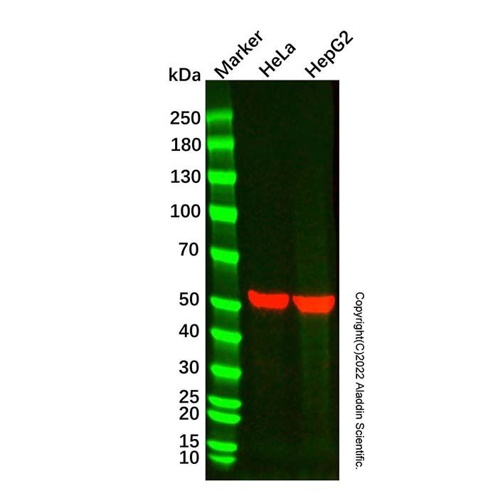
Caspase-9 Mouse mAb (Ab183845) - Western Blot All lanes: Caspase-9 Mouse Antibody (Ab183845) at 1/10000 dilution Samples: Lysates at 20 µg per lane Secondary: Goat Anti-Mouse IgG H&L (HRP) (Ab179001) at 1/20000 dilution
Predicted band size: 46 kDa Observed band size: 52 kDa Exposure time: 10.0 s
Caspase-9 Mouse mAb (Ab183845) - Western Blot All lanes: Caspase-9 Mouse Antibody (Ab183845) at 1/10000 dilution Samples: Lysates at 20 µg per lane Secondary: Goat Anti-Mouse IgG H&L (HRP) (Ab179001) at 1/20000 dilution
Predicted band size: 46 kDa Observed band size: 53 kDa Exposure time: 15.0 s
Caspase-9 Mouse mAb (Ab183845) - Flow Cytometry Flow Cytometry analysis of K562 cells stained with Caspase-9 Mouse mAb (Ab183845) (Red) at 5 μg/mL for 1 hour at 4°C. Cells were fixed and permeabilized with 100% methanol. The Goat Anti-Mouse IgG H&L (PE) (Ab179006) was used as the secondary antibody. Unlabeled sample (Black) was used as a negative control. Mouse IgG2b-kappa, anti-E8L (Ab210600) was used as an isotype control (Blue).
Caspase-9 Mouse mAb (Ab183845) - IF/ICC IF analysis of Caspase-9 (Green) in HeLa cells. The cells were fixed and permeabilized with 100% methanol for 5 minutes, and blocked with 2% BSA for 1 hour at room temperature. Cells were stained with Caspase-9 Mouse mAb (Ab183845) at 2.0 μg/mL in blocking buffer for 2 hour at RT and then incubated with Goat Anti-Mouse IgG H&L (FITC) (Ab179003) at a dilution of 1/1000 for 1 hour at room temperature in the dark. Cells were counterstained with DAPI (Blue). Images were taken on the confocal laser scanning microscope.

 ZJ25F0622493


 危险品化学品经营许可证(带存储)