Calcium-sensitive protein which reversibly blocks the barbed ends of actin filaments but does not sever preformed actin filaments. May play an important role in macrophage function. May play a role in regulating cytoplasmic and/or nuclear structures through potential interactions with actin. May bind DNA.
宿主种属
小鼠(Mouse)
特异性
Actin Regulatory Protein CAPG/MCP
种属反应性
人(Human),大鼠(Rat),小鼠(Mouse)
免疫原
Recombinant protein of human Actin Regulatory Protein CAPG/MCP (AA 191-270).
阳性对照
WB: THP-1 and U937cell lysates. IHC: Human lung tissue.
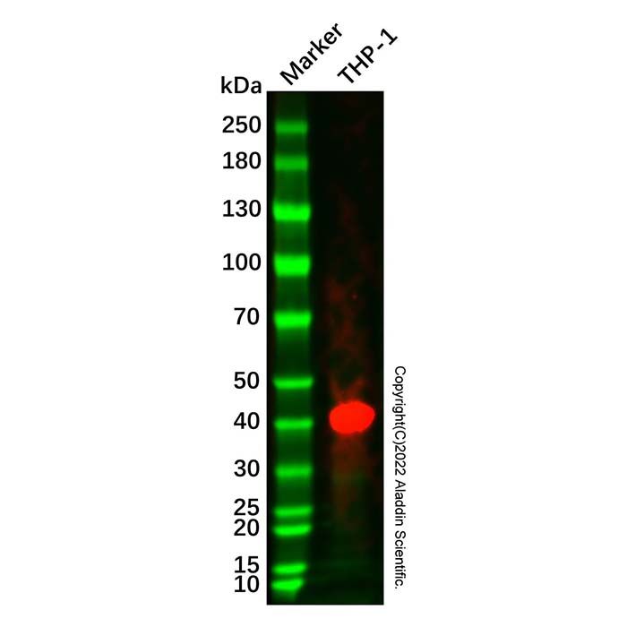
Actin Regulatory Protein CAPG/MCP Mouse mAb (Ab334429) - Western Blot
All lanes: Actin Regulatory Protein CAPG/MCP Mouse mAb (Ab334429) at 1/1000 dilution
Samples: Lysates at 20 µg per lane
Secondary: Goat Anti-Mouse IgG H&L (HRP) (Ab179001) at 1/20000 dilution
Predicted band size: 38 kDa
Observed band size: 41 kDa
Exposure time: 44.1 s
Actin Regulatory Protein CAPG/MCP Mouse mAb (Ab334429) - Western Blot
All lanes: Actin Regulatory Protein CAPG/MCP Mouse mAb (Ab334429) at 1/1000 dilution
Samples: Lysates at 20 µg per lane
Secondary: Goat Anti-Mouse IgG H&L (HRP) (Ab179001) at 1/20000 dilution
Predicted band size: 38 kDa
Observed band size: 42 kDa
Exposure time: 44.1 s
Actin Regulatory Protein CAPG/MCP Mouse mAb (Ab334429) - IHC
IHC-P was performed using sections of the formalin-fixed paraffin-embedded lung tissue. Antigen was retrieved through addition of boiling Tris/EDTA buffer pH 9 in a pressure cooker for 3 min. Endogenous peroxidase activity was quenched by incubating the sections with 3% H2O2 for 30 min at room temperature. The sections were then incubated with Actin Regulatory Protein CAPG/MCP Mouse mAb (Ab334429) at room temperature for 1 h. Poly-peroxidase conjugated goat anti-mouse IgG was used as the secondary antibody. Diaminobenzidine was used as the chromogen. The section was counterstained with hematoxylin. A tissue section incubated with phosphate-buffered saline followed by incubation with the secondary antibody was used as the background control.
Result: Macrophages are positively stained at the Cytoplasm nuclei and cell membrane.

 ZJ26F0435057


 危险品化学品经营许可证(带存储)