葡萄糖(Glucose),被认为是自然界中分布最广且最为重要的一种单糖,可以被直接吸收并进入血液,是人体能量代谢和维持生命所必需的营养物质之一,对维持机体代谢与内环境稳态有非常重要的作用。天然的葡萄糖均为右旋即D型,即D-Glucose。葡萄糖是生物体重要的能量来源和代谢中间产物。葡萄糖一方面是光合作用的主要产物,另一方面也是呼吸反应的主要底物。在呼吸反应中,葡萄糖通过一系列酶促反应氧化生成二氧化碳和水,同时生成重要的能量分子ATP。由于葡萄糖在能量代谢方面的重要性,血糖水平已经成为多种代谢疾病的重要诊断指标。病理性高血糖与许多疾病相关,如糖尿病、甲亢以及垂体和肾上腺亢进,而病理性低血糖常见于胰岛素分泌相关的肿瘤、黏液性水肿、垂体及肾上腺功能减退症等疾病。同时细胞内的葡萄糖水平也是监测细胞代谢状态的一个重要指标。
本试剂盒的检测原理为葡萄糖在葡萄糖氧化酶(GOD)的催化作用下和氧气发生反应,生成D-葡萄糖酸(Gluconic acid)和H2O2。生成的H2O2在过氧化物酶(Peroxidase, POD)的作用下与苯酚(Phenol)和4-氨基安替比林(4-Aminoantipyrine)生成红色的醌亚胺(Quinoneimine),再通过检测红色醌亚胺在510nm的吸光度来最终计算样品中葡萄糖的含量。吸光度与样品中葡萄糖的含量成正比。
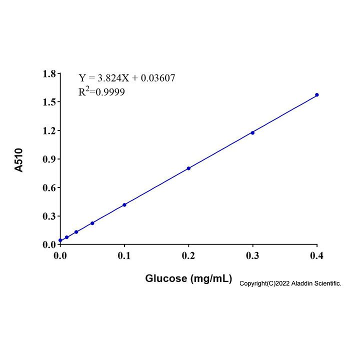
本试剂盒检测灵敏度高,线性范围宽,样品用量少。在样品体积为40 µL时,可以检测浓度低至0.01 mg/mL的葡萄糖,在0.01-0.4 mg/mL(即0.056-2.22 mM)浓度范围内有良好的线性关系。
使用方法
1. 样本制备
(1)血液样品的准备:对于血清样品,将全血在常温下静置30分钟-2小时,4ºC约1000-2000×g离心10分钟,取上清;对于血浆样品,将全血用肝素或者EDTA进行抗凝,4ºC约1000-2000×g离心10分钟,取上清。
(2)细胞样本的准备:对于培养的贴壁细胞,PBS洗涤细胞一次。对于培养的悬浮细胞,离心收集细胞用PBS洗涤细胞一次。按照每100万细胞加入200 μL 裂解液的比例加入裂解液,适当吹打,冰浴10分钟充分裂解细胞。4ºC, 12,000×g离心5分钟,取上清用于后续检测。
(3)对于组织样品,按照每10 mg组织加入100 μL 裂解液的比例,使用匀浆器冰浴匀浆。4ºC约12,000×g离心5分钟,取上清用于后续检测。
(4)细胞培养上清样品的准备:对于贴壁细胞,直接取培养液;对于悬浮细胞,离心取培养液。
以上所有操作均需在4ºC或冰上操作。制备好的细胞或组织样品如果不能立即检测,可以-20ºC或-80ºC冻存。
2. 标准曲线的设置
取30 μL 葡萄糖标准品 (4 mg/mL),加入270 μL 裂解液或检测缓冲液(如果检测的是用裂解液制备的细胞或组织裂解样品,使用裂解液;如果检测的是血液、上清等无需裂解处理的样品,使用检测缓冲液),配制成浓度为0.4 mg/mL的葡萄糖标准溶液。分别取0.4 mg/mL的葡萄糖标准溶液0、1、2.5、5、10、20、30、40 μL加入96孔板的标准品孔中,并相应地用裂解液或检测缓冲液补足至40 μL,此时,标准曲线的浓度分别为0、0.01、0.025、0.05、0.1、0.2、0.3、0.4 mg/mL。
3. 显色工作液的配制
按照每个检测反应160 μL的体积配制显色工作液。根据待检测样品(包括标准品)的数量,配制适量的显色工作液。具体配制方法参考下表,建议尽量现配现用。
样本数 | 1 | 10 | 20 | 50 | 检测缓冲液 | 132 μL | 1320 μL | 2640 μL | 6600 μL | 酶液 | 2 μL | 20 μL | 40 μL | 100 μL | 检测试剂A | 8 μL | 80 μL | 160 μL | 400 μL | 检测试剂B | 18 μL | 180 μL | 360 μL | 900 μL | 总体积 | 160 μL | 1600 μL | 3200 μL | 8000 μL |
注:H2O2的存在会对葡萄糖的检测产生干扰。如果样品含有H2O2,须同时设置样品背景对照孔,加入不含酶液的显色工作液,即配制显色工作液时2 µL 酶液用检测缓冲液替代。计算时样品孔的读数需要减去样品背景对照孔的读数。
|
4. 样本测定
(1)取1-40 μL样品或稀释后的样品至96孔板样品孔中,并相应地加入裂解液或检测液补足至40 µL。同时设置仅含裂解液或检测缓冲液的孔为空白对照孔(如果检测需要裂解处理的细胞或组织样品,使用裂解液;如果检测血液、上清等无需裂解处理的样品,使用检测缓冲液)。
(2)各孔加入显色工作液160 μL,混匀。37ºC避光反应15分钟。
(3)反应结束后,请在A510nm进行吸光度检测。
5. 结果计算
建立标准曲线,并计算样品中葡萄糖的浓度(A)。葡萄糖浓度的计算公式如下:C (mg/ml)=A×n
注:A为根据标准曲线确定的葡萄糖含量(mg/mL);n为样品总稀释倍数。可根据葡萄糖的分子量180计算出样品中葡萄糖的摩尔浓度(mM)=C/0.18。
注意事项
1. 建议尽量现配现用显色工作液。
2. H2O2的存在会对葡萄糖的检测产生干扰。如果样品含有H2O2,须同时设置样品背景对照孔,加入不含酶液的显色工作液,即配制显色工作液时2 µL 酶液用检测缓冲液替代。计算时样品孔的读数需要减去样品背景对照孔的读数。
3. 血清等样品如在4ºC保存,保存时间不得超过2周,否则会影响检测结果的准确性。通常血清样品宜-20ºC保存,-80ºC保存更佳。
Glucose, considered one of the most widely distributed and important monosaccharides in nature, can be directly absorbed into the bloodstream. It is an essential nutrient for human energy metabolism and life maintenance, playing a critical role in sustaining metabolism and homeostasis. Naturally occurring glucose exists exclusively in the dextrorotatory (D) form, namely D-Glucose. Glucose serves as a major energy source and a key metabolic intermediate in living organisms. On one hand, it is a primary product of photosynthesis; on the other, it is a major substrate in respiration. During respiration, glucose is oxidized through a series of enzymatic reactions to carbon dioxide and water, while generating the crucial energy molecule ATP. Due to its central role in energy metabolism, blood glucose levels have become an important diagnostic marker for various metabolic diseases. Pathological hyperglycemia is associated with conditions such as diabetes mellitus, hyperthyroidism, and pituitary or adrenal hyperactivity, whereas pathological hypoglycemia is commonly observed in tumors related to insulin secretion, myxedema, and pituitary or adrenal insufficiency. Additionally, intracellular glucose levels serve as an important indicator for monitoring cellular metabolic status.
The detection principle of this kit is as follows: Glucose is oxidized by glucose oxidase (GOD) in the presence of oxygen to produce D-gluconic acid and H₂O₂. The generated H₂O₂ then reacts with phenol and 4-aminoantipyrine under the catalysis of peroxidase (POD) to form a red quinoneimine chromophore. The absorbance of the red quinoneimine at 510 nm is measured to determine the glucose concentration in the sample. The absorbance is directly proportional to the amount of glucose present.
This assay kit demonstrates high detection sensitivity, a wide linear range, and low sample consumption. With a sample volume of 40 µL, it can detect glucose concentrations as low as 0.01 mg/mL and exhibits a strong linear relationship within the range of 0.01–0.4 mg/mL (equivalent to 0.056–2.22 mM).
Usage Protocol
1. Sample Preparation
1) Preparation of Blood Samples:
For serum samples: Allow whole blood to clot at room temperature for 30 minutes to 2 hours. Centrifuge at approximately 1000-2000 × g for 10 minutes at 4°C and collect the supernatant.
For plasma samples: Collect whole blood using heparin or EDTA as an anticoagulant. Centrifuge at approximately 1000-2000 × g for 10 minutes at 4°C and collect the supernatant.
2) Preparation of Cell Samples:
For cultured adherent cells: Wash the cells once with PBS; For cultured suspension cells: Centrifuge to collect the cells and wash them once with PBS. Add Lysis Buffer at a ratio of 200 µL per 1 million cells. Pipette appropriately to mix and incubate on ice for 10 minutes for complete lysis. Centrifuge at 12,000 × g for 5 minutes at 4°C. Collect the supernatant for subsequent assays.
3) Preparation of Tissue Samples:
Add Lysis Buffer at a ratio of 100 µL per 10 mg of tissue. Homogenize the tissue using a homogenizer on ice. Centrifuge at approximately 12,000 × g for 5 minutes at 4°C. Collect the supernatant for subsequent assays.
4) Preparation of Cell Culture Supernatant Samples:
For adherent cells: Directly collect the culture medium; For suspension cells: Centrifuge the culture and collect the supernatant.
All the above procedures must be performed at 4°C or on ice. Prepared cell or tissue samples can be stored at -20°C or -80°C if not assayed immediately.
2. Establishment of Standard Curve
Take 30 μL of the Glucose Standard (4 mg/mL) and add it to 270 μL of lysis buffer or assay buffer (use lysis buffer for lysed cell or tissue samples prepared with lysis buffer; use assay buffer for samples such as blood or supernatant that do not require lysis treatment), to prepare a 0.4 mg/mL glucose standard solution. Then pipette 0, 1, 2.5, 5, 10, 20, 30, and 40 μL of the 0.4 mg/mL glucose standard solution into the standard wells of a 96‑well plate, and bring the volume in each well to 40 μL by adding the corresponding lysis buffer or assay buffer. The resulting standard curve concentrations are 0, 0.01, 0.025, 0.05, 0.1, 0.2, 0.3, and 0.4 mg/mL, respectively.
3. Preparation of Chromogenic Solution
Prepare the Chromogenic Solution based on a volume of 160 μL per reaction. Prepare an appropriate amount according to the number of samples to be tested (including standards). For specific preparation methods, refer to the table below. It is recommended to prepare the working solution freshly before use.
Samples | 1 | 10 | 20 | 50 | Assay Buffer | 132 μL | 1320 μL | 2640 μL | 6600 μL | Enzyme Solution | 2 μL | 20 μL | 40 μL | 100 μL | Assay Reagent A | 8 μL | 80 μL | 160 μL | 400 μL | Assay Reagent B | 18 μL | 180 μL | 360 μL | 900 μL | Total volume | 160 μL | 1600 μL | 3200 μL | 8000 μL |
Note: The presence of H₂O₂ may interfere with the glucose detection. If the sample contains H₂O₂, a sample background control well must be set up in parallel. Add the Chromogenic Solution without the enzyme solution (replace the 2 μL of enzyme solution with Assay Buffer). When calculating results, subtract the absorbance reading of the sample background control well from that of the sample well. |
4. Sample Assay
1) Add 1–40 μL of sample or diluted sample to the sample wells of a 96‑well plate. Then add the corresponding Lysis Buffer or Assay Buffer to bring the volume to 40 μL. At the same time, set up wells containing only Lysis Buffer or Assay Buffer as blank control wells (use Lysis Buffer if detecting cell or tissue samples that require lysis; use Assay Buffer if detecting samples such as blood or supernatant that do not require lysis).
2) Add 160 μL of Chromogenic Solution to each well and mix. Incubate at 37 °C in the dark for 15 minutes.
3) After the reaction, measure the absorbance at 510 nm (A₅₁₀).
5. Calculation of Results
Establish the standard curve and calculate the glucose concentration in the sample (A). The formula for calculating glucose concentration is as follows: C (mg/mL) = A × n
Note: A is the glucose concentration (mg/mL) determined from the standard curve; n is the total dilution factor of the sample.
The molar concentration of glucose in the sample (mM) can be calculated using the molecular weight of glucose (180): Glucose molar concentration (mM) = C / 0.18.
Precautions
1. It is recommended to prepare the Color Developer Working Solution freshly before use whenever possible.
2. The presence of H₂O₂ may interfere with glucose detection. If the sample contains H₂O₂, a sample background control well must be set up. Add the Color Developer Working Solution prepared without the enzyme solution (replace the 2 μL of enzyme solution with Assay Buffer). When calculating results, subtract the absorbance reading of the sample background control well from that of the sample well.
3. If serum or similar samples are stored at 4 °C, the storage time should not exceed two weeks; otherwise, the accuracy of the detection results may be affected. Generally, serum samples are recommended to be stored at –20 °C, and storage at –80 °C is even better.

 ZJ26F0737076

 危险品化学品经营许可证(带存储)