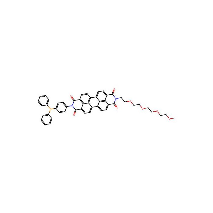
Liperfluo是Spy-LHP的类似物,可用于检测脂质过氧化物 (LPO),Liperfluo会被脂质过氧化物特异性氧化而在乙醇等有机溶剂中发出很强的荧光 (图1,2)。由于氧化型Liperfluo的激发和发射波长分别为524 nm和 535 nm (均为长波长),因此可以减轻光对测定样品的损伤和降低样品的自发荧光。由于Liperfluo在二异喹啉环的一个末端连接有四乙二醇基团,因此提高了它在水溶液中的分子分散性。虽然它的氧化型在水溶液中几乎没有荧光,但是在细胞膜等脂溶性高的部位会产生较强的荧光。因此Liperfluo适用于活细胞中脂质过氧化物的荧光成像或流式分析。

特点:
(1)能够成像和检测细胞中的脂质过氧化物;
(2)长波长激发,可以减少光损伤和自发荧光对细胞的影响;
(3)高度脂质过氧化物特异性。
研究领域
在铁死亡研究中:作为细胞死亡形式之一,在铁死亡研究中使用到Liperfluo。
溶液配制
1、配制 1 mmol/L Liperfluo DMSO 储存液
在含有 50 μg Liperfluo 的管子中加入 60 μl 新鲜无水的 DMSO,用移液器反复吹打,最后用涡旋振荡器振荡直至肉眼观察完全溶解,制备成 1 mmol/l 的 Liperfluo DMSO 储存液。
2、配制Liperfluo工作液
最佳染色的Liperfluo浓度将根据实验条件而变化,用于荧光显微成像时,遵循下列步骤:
用于荧光成像:用无血清培养基稀释 1 mmol/L Liperfluo DMSO 储存液,调整到合适的浓度(1-20 μmol/l)制备成 Liperfluo工作液。
用于流式检测:将 1 mmol/L Liperfluo DMSO储存液直接加入待染色的细胞悬浮液中。
※Liperfluo 管子的管壁上可能会有粉末残留,请用 DMSO 溶液反复吹洗,直至粉末完全被洗脱下来并溶解。
※由于 Liperfluo 见光易分解,因此配制好的 Liperfluo 溶液需用铝箔纸包裹并在 1 天内用完。
※如果 Liperfluo 无法完全溶解,请在避光的同时用超声或用 40℃水浴加热 3 min 的方法帮助溶解。
※采用超声处理时,请按下图所示,在管子底部注水排除空气后再进行超声。

*1 Liperfluo 对光敏感,应在 1 分钟内完成移液。
*2 如 Liperfluo 固体在管体底部,在吹打过程中,使用枪头将大块固体打成小块。
*3 如涡旋振荡后固体仍存在,超声或加热 (40 ℃,30 min) 管体,以达到溶解目的。超声时请注意管体下部填充水。 详见下图。

2、配制1 mmol/l的Liperfluo DMSO stock solution
最佳染色的Liperfluo浓度将根据实验条件而变化,用于荧光显微成像时,遵循下列步骤:
用于荧光成像:用无血清培养基稀释1mmol/l Liperfluo DMSO溶液,制备成工作溶液。
用于流式检测:将1 mmol/l Liperfluo DMSO溶液直接加入待染色的细胞悬浮液中。
操作步骤
细胞中脂质过氧化物的检测
1.在培养皿或孔板中接种细胞。
2.去除上清液,用无血清培养基洗涤细胞1次。
3.加入适量的Liperfluo工作液,37℃孵育30 min。
*根据实验条件等不同因素,Liperfluo的最佳浓度也不同。需要提前摸索条件。
4.去除上清液并用无血清培养基洗涤细胞2次。
5.在荧光显微镜下观察细胞或使用流式细胞仪分析细胞。
荧光谱图
Liperfluo is an analog of Spy‑LHP and can be used for the detection of lipid peroxides (LPO). Liperfluo is specifically oxidized by lipid peroxides and emits strong fluorescence in organic solvents such as ethanol (Figures 1, 2). The oxidized Liperfluo exhibits excitation and emission wavelengths at 524 nm and 535 nm, respectively (both long‑wavelength), which reduces photodamage to test samples and decreases sample autofluorescence. Bearing a tetraethylene glycol moiety attached to one terminus of the diisoquinoline ring, Liperfluo possesses improved molecular dispersibility in aqueous solution. Although its oxidized form shows barely any fluorescence in water, it generates intense fluorescence in lipid‑rich compartments such as cell membranes. Accordingly, Liperfluo is suitable for fluorescence imaging or flow cytometric analysis of lipid peroxides in living cells.

Features
1. Enables imaging and detection of lipid peroxides in cells;
2. Long‑wavelength excitation mitigates cellular photodamage and interference from autofluorescence;
3. High specificity toward lipid peroxides.
Research Areas
Ferroptosis research: Liperfluo is applied in the study of ferroptosis, one form of regulated cell death.
Solution Preparation
1. Preparation of 1 mmol/L Liperfluo DMSO Stock Solution
Add 60 μL of fresh anhydrous DMSO to the vial containing 50 μg Liperfluo. Pipette up and down repeatedly, then vortex using a vortex mixer until complete dissolution is observed visually, to prepare the 1 mmol/L Liperfluo DMSO stock solution.
2. Preparation of Liperfluo Working Solution
The optimal Liperfluo concentration for staining varies depending on experimental conditions. Follow the steps below for fluorescence microscopy imaging:
For fluorescence imaging: Dilute the 1 mmol/L Liperfluo DMSO stock solution with serum-free medium to an appropriate concentration (1–20 μmol/L) to prepare the Liperfluo working solution.
For flow cytometry detection: Add the 1 mmol/L Liperfluo DMSO stock solution directly into the cell suspension to be stained.
※ Powder residue may remain on the inner wall of the Liperfluo vial. Rinse repeatedly with DMSO until all powder is washed off and dissolved.
※ Liperfluo is light-sensitive and readily decomposes upon exposure to light. Wrap the prepared Liperfluo solution with aluminum foil and use it within 1 day.
※ If Liperfluo fails to dissolve completely, perform the operation under light protection. Sonication or heating in a 40 ℃ water bath for 3 min can be applied to aid dissolution.
※ For sonication, add water to the bottom of the tube to remove air before sonication, as shown in the figure below.

*1 Liperfluo is light‑sensitive; perform pipetting within 1 minute.
*2 If solid Liperfluo adheres to the bottom of the vial, crush large solid chunks into small pieces with the pipette tip during pipetting.
*3 If solids remain after vortexing, sonicate or heat the vial (40 °C, 30 min) to achieve full dissolution. Ensure the lower portion of the vial is submerged in water during sonication. See the figure below for details.

2. Preparation of 1 mmol/L Liperfluo DMSO Stock Solution
The optimal Liperfluo concentration for staining varies with experimental conditions. Follow the procedures below for fluorescence microscopy:
Fluorescence imaging: Dilute the 1 mmol/L Liperfluo DMSO stock with serum‑free medium to prepare the working solution.
Flow cytometry: Add the 1 mmol/L Liperfluo DMSO stock directly into the cell suspension to be stained.
Operating Procedure
Detection of lipid peroxides in cells
1. Seed cells in culture dishes or well plates.
2. Discard supernatant and wash cells once with serum‑free medium.
3. Add an appropriate volume of Liperfluo working solution, and incubate at 37 °C for 30 min. *The optimal Liperfluo concentration depends on experimental conditions; condition optimization in advance is required.
4. Remove supernatant and wash cells twice with serum‑free medium.
5. Observe cells under a fluorescence microscope or analyze cells by flow cytometry.
Fluorescence spectra




 危险品化学品经营许可证(带存储)