还原糖(RS)广泛存在于动物、植物、微生物和培养细胞中。植物体内的还原糖主要包括葡萄糖、果糖和麦芽糖等,其中葡萄糖和果糖不仅是呼吸作用的主要底物,也是进一步合成蔗糖、淀粉和纤维素的底物。
检测原理:在碱性溶液中3,5-二硝基水杨酸能够被RS还原生成棕红色的氨基化合物,在540nm处有特征吸收峰,在一定的浓度范围内,RS含量与540nm吸光度成线性关系,根据标准曲线,即可计算出样品中RS的含量。
检测范围:0.05-0.6 mg/mL
灵敏度:0.025 mg/mL
适用样本:植物组织、动物组织、细胞、细菌、血清(浆)
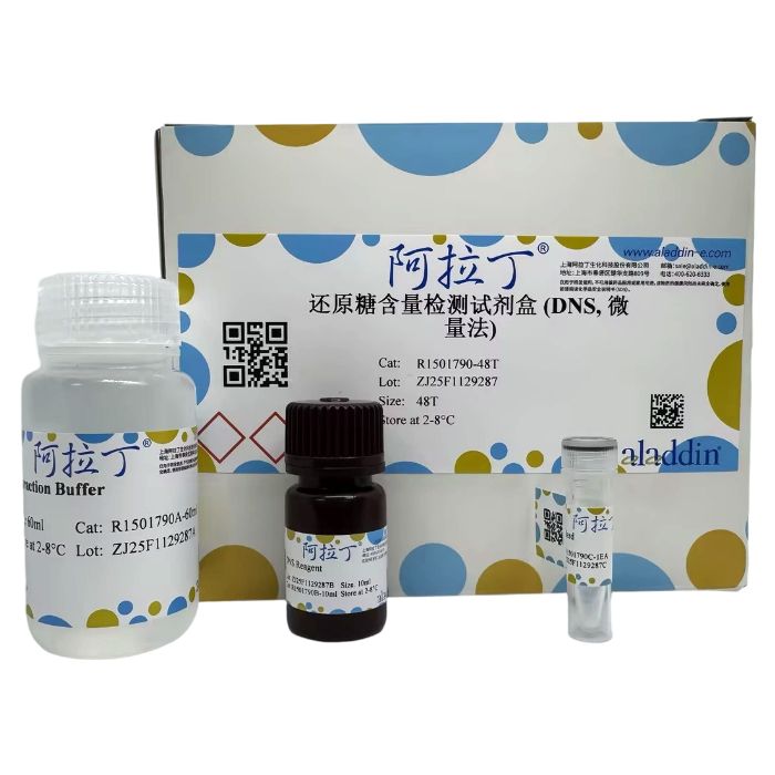
R1501790
| Component
| 48T
| 96T
| Storage
|
R1501790A
| Extraction Buffer
| 60 mL
| 120 mL
| 2-8℃
|
R1501790B
| DNS Reagent
| 10 mL
| 20 mL
| 2-8℃. Store in the dark.
|
R1501790C
| Standard
| 1EA
| 1EA
| 2-8℃ |
注意:正式检测前,建议选择2-3个预期差异较大的样本进行预实验。
自备仪器和试剂
酶标仪或可见分光光度计(能测540 nm处的吸光度)
96孔板或微量玻璃比色皿,可调节式移液枪及枪头
离心机,水浴锅
去离子水
匀浆器(如果是组织样本)
实验流程
1、试剂准备
试剂名称
| 试剂准备
| 注意事项
|
Extraction Buffer
| 即用型;使用前平衡到室温
| 4℃保存;有一定的刺激性,建议在使用过程中做好个人防护
|
DNS Reagent
| 即用型;使用前平衡到室温
| 4℃避光保存;有一定的刺激性,建议在使用过程中做好个人防护
|
Standard
| 临用前加入1mL去离子水溶解,配制10 mg/mL标准品
| 4℃保存2周 |
2、标准曲线设置
用去离子水将10 mg/mL标准品稀释为0.6,0.5,0.4,0.3,0.2,0.1,0.05 mg/mL。
| 序号 | 10 mg/mL标准品体积 (μL)
| 去离子水体积 (μL)
| 浓度 (mg/mL)
|
| Std.1 | 60 | 940 | 0.6 |
Std.2
| 50 | 950 | 0.5 |
Std.3
| 40 | 960 | 0.4 |
Std.4
| 30 | 970 | 0.3 |
Std.5
| 20 | 980 | 0.2 |
Std.6
| 10 | 990 | 0.1 |
Std.7
| 5 | 995 | 0.05 |
注意:每次实验都要做一次标准品检测,制作标曲;稀释后的标准品溶液不稳定,必须在4小时内使用。
3、样本制备
3.1 植物或动物组织样本
称取约0.1g组织,加入1mL Extraction Buffer,冰浴匀浆,转移到有盖离心管中(防止加热时水分蒸发),80℃水浴40min,每5min振摇混匀1次,8,000 g,25°C离心10min,取上清液待测。
3.2 细菌或细胞
收集细菌或细胞到离心管内,弃上清;按照每500万细菌或细胞加入1mL Extraction Buffer,冰浴超声波破碎细菌或细胞5min(功率20%,超声3s,间隔10s,重复30次),转移到有盖离心管中(防止加热时水分蒸发),80°C水浴40min,每5min振摇混匀1次,8,000 g,25°C离心10min,取上清液待测。
3.3 血清(浆)样本
取0.1mL血清(浆),加0.9mL Extraction Buffer,充分混匀;转移到有盖离心管中(防止加热时水分蒸发),80℃水浴40min,每5min振摇混匀1次,8,000 g,25°C离心10min,取上清液待测。
注意:
如需测定蛋白浓度,推荐使用阿拉丁B665595 BCA蛋白定量试剂盒或R1491648 即用型BCA蛋白定量试剂盒。Extraction Buffer中含有使蛋白变性的成分,若按蛋白浓度计算时,需要重新提取蛋白进行测定。
4、实验步骤
4.1 酶标仪或可见分光光度计预热30min以上,调节波长到540nm,可见分光光度计用去离子水调零。
4.2 操作表
| 试剂 | 空白管 (μL) | 标准管 (μL)
| 测定管 (μL)
| 对照管 (μL)
|
| Sample | 0 | 0
| 175
| 175
|
| 不同浓度Std. | 0
| 175
| 0
| 0
|
| 去离子水 | 175 | 0
| 0
| 125
|
DNS Reagent
| 125 | 125
| 125
| 0 |
混匀,沸水浴加热5min(盖紧,防止水分蒸发),取出后立即冷却至室温。取200μL至96孔板或微量玻璃比色皿中,540nm波长处测定吸光值。计算ΔA 测定 =A 测定 -A 对照 ,ΔA 标准 =A 标准 -A 空白 。
注意:
空白管和标准管只需做1-2次。
实验之前建议选择2-3个预期差异大的样本做预实验。如果ΔA 测定 小于0.04可适当加大样本量。如果ΔA 测定 大于0.6 mg/mL的ΔA 标准 ,样本可用Extraction Buffer进一步稀释,计算结果乘以稀释倍数,或减少提取用样本量。
5、结果计算
注意:我们为您提供的计算公式,包括推导过程计算公式和简洁计算公式。两者完全相等。建议以加粗的简洁计算公式为最终计算公式。
5.1 标准曲线的绘制
以标准品浓度为y轴,ΔA 标准 为x轴,绘制标准曲线。将ΔA 测定 带入方程计算出y值。
5.2 样本还原糖含量计算
(1) 按样本质量计算
还原糖 (μg/g)=1000×y×V 提取 ÷W×n=1000×y÷W×n
(2) 按样本蛋白浓度计算
还原糖 (μg/mg prot)=1000×y×V 提取 ÷(V 提取 ×Cpr)×n=1000×y÷Cpr×n
(3) 按细菌或细胞数量计算
还原糖 (μg/10⁴)=1000×y×V 提取 ÷500×n=2×y×n
(4) 按血清(浆)体积计算
还原糖 (μg/mL)=1000×y×V 提取 ÷V 液 ×n=10000×y×n
参数说明
1000:单位换算系数,1 mg/mL=1000 μg/mL;
V 提取 :加入Extraction Buffer体积,1 mL;
V 液 :加入血清(浆)体积,0.1 mL;
Cpr:样本蛋白质浓度,mg/mL;
W:样本质量,g;
500:细菌或细胞总数,500万;
n:稀释倍数
6、结果展示
典型标准曲线:y=0.2243x+0.0545,R²=0.9957

注意事项
本产品仅供科学研究使用,不适用于临床诊断。为了您的安全和健康,请穿实验服并戴一次性手套操作。
Reducing sugars (RS) are widely present in animals, plants, microorganisms, and cultured cells. Reducing sugars in plants primarily include glucose, fructose, and maltose. Among these, glucose and fructose are not only the main substrates for respiration but also serve as substrates for the further synthesis of sucrose, starch, and cellulose.
Detection Principle: In an alkaline solution, 3,5-dinitrosalicylic acid (DNS) can be reduced by reducing sugars to produce a brown-red-colored amino compound, which has a characteristic absorption peak at 540 nm. Within a certain concentration range, the RS content is linearly correlated with the absorbance at 540 nm. The RS content in the sample can be calculated based on a standard curve.
Detection Range: 0.05 - 0.6 mg/mL
Sensitivity: 0.025 mg/mL
Applicable Samples: Plant tissues, animal tissues, cells, bacteria, serum (plasma)
R1501790
| Component
| 48T
| 96T
| Storage
|
R1501790A
| Extraction Buffer
| 60 mL
| 120 mL
| 2-8℃
|
R1501790B
| DNS Reagent
| 10 mL
| 20 mL
| 2-8℃. Store in the dark.
|
R1501790C
| Standard
| 1EA
| 1EA
| 2-8℃ |
Note: Before formal testing, it is recommended to perform a preliminary test with 2-3 samples expected to have significant differences.
User-Prepared Instruments and Reagents
Microplate reader or visible spectrophotometer (capable of measuring absorbance at 540 nm)
96-well plate or micro glass cuvettes, adjustable micropipettes and tips
Centrifuge, water bath
Deionized water
Homogenizer (for tissue samples)
Experimental Procedure
1. Reagent Preparation
Reagent Name
| Reagent Preparation
| Notes
|
Extraction Buffer
| Ready-to-use; Equilibrate to room temperature before use.
| Store at 4°C. Slightly irritating. Use appropriate personal protective equipment.
|
DNS Reagent
| Ready-to-use; Equilibrate to room temperature before use.
| Store at 4°C protected from light. Slightly irritating. Use appropriate personal protective equipment.
|
Standard
| Before use, add 1 mL of deionized water to dissolve, preparing a 10 mg/mL stock standard solution.
| Can be stored at 4°C for 2 weeks. |
2. Standard Curve Setup
Dilute the 10 mg/mL standard stock solution with deionized water to concentrations of 0.6, 0.5, 0.4, 0.3, 0.2, 0.1, and 0.05 mg/mL.
| Tube | Volume of 10 mg/mL Standard (μL) | Volume of Deionized Water (μL)
| Concentration (mg/mL)
|
| Std.1 | 60 | 940 | 0.6 |
Std.2
| 50 | 950 | 0.5 |
Std.3
| 40 | 960 | 0.4 |
Std.4
| 30 | 970 | 0.3 |
Std.5
| 20 | 980 | 0.2 |
Std.6
| 10 | 990 | 0.1 |
Std.7
| 5 | 995 | 0.05 |
Note: The standard curve must be generated with each experiment. Diluted standard solutions are unstable and must be used within 4 hours.
3. Sample Preparation
3.1 Plant or Animal Tissue Samples
Weigh approximately 0.1 g of tissue. Add 1 mL of Extraction Buffer and homogenize in an ice bath. Transfer the homogenate to a capped centrifuge tube (to prevent evaporation during heating). Incubate in an 80°C water bath for 40 minutes, vortexing every 5 minutes. Centrifuge at 8,000 g, 25°C for 10 minutes. Collect the supernatant for assay.
3.2 Bacteria or Cells
Collect bacteria or cells into a centrifuge tube; discard the supernatant. Add 1 mL of Extraction Buffer per 5 million bacteria/cells. Sonicate in an ice bath for 5 minutes (power 20%, pulse 3s on, 10s off, repeat 30 times). Transfer to a capped centrifuge tube (to prevent evaporation during heating). Incubate in an 80°C water bath for 40 minutes, vortexing every 5 minutes. Centrifuge at 8,000 g, 25°C for 10 minutes. Collect the supernatant for assay.
3.3 Serum (Plasma) Samples
Take 0.1 mL of serum (plasma) and add 0.9 mL of Extraction Buffer; mix thoroughly. Transfer to a capped centrifuge tube (to prevent evaporation during heating). Incubate in an 80°C water bath for 40 minutes, vortexing every 5 minutes. Centrifuge at 8,000 g, 25°C for 10 minutes. Collect the supernatant for assay.
Note:
If protein concentration measurement is required, Aladdin's BCA Protein Quantification Kit (B665595) or Ready-to-Use BCA Protein Quantification Kit (R1491648) is recommended. The Extraction Buffer contains components that denature proteins. If calculating based on protein concentration, protein needs to be re-extracted separately for measurement.
4. Assay Steps
4.1 Preheat the microplate reader or visible spectrophotometer for at least 30 minutes. Set the wavelength to 540 nm. For spectrophotometers, zero the instrument with deionized water.
4.2 Assay Procedure:
| Reagent | Blank Tube (μL) | Standard Tube (μL)
| Test Tube (μL)
| Control Tube (μL)
|
| Sample | 0 | 0
| 175
| 175
|
| Standard (various conc.) | 0
| 175
| 0
| 0
|
| Deionized Water | 175 | 0
| 0
| 125
|
DNS Reagent
| 125 | 125
| 125
| 0 |
Mix well. Heat in a boiling water bath for 5 minutes (cap tightly to prevent evaporation). Remove and immediately cool to room temperature. Transfer 200 μL to a 96-well plate or micro glass cuvette. Measure the absorbance at 540 nm. Calculate ΔA test = A test - A control , ΔA standard = A standard - A blank .
Note:
The Blank and Standard tubes only need to be set up 1-2 times.
It is recommended to perform a preliminary test with 2-3 samples expected to have significant differences before the formal experiment. If ΔA <sub> test </sub> is less than 0.04, consider increasing the sample volume appropriately. If ΔA <sub> test </sub> is greater than the ΔA <sub> standard </sub> of the 0.6 mg/mL standard, further dilute the sample with Extraction Buffer (multiply the result by the dilution factor) or reduce the amount of sample used for extraction.
5. Calculation of Results
Note: We provide both the derived formula and a simplified formula. They are equivalent. It is recommended to use the simplified formula in bold for final calculation.
5.1 Standard Curve Plotting
Plot the standard concentration (y-axis) against ΔA standard (x-axis) to generate the standard curve. Substitute ΔA test into the standard curve equation to calculate y (mg/mL).
5.2 Sample Reducing Sugar Content Calculation
(1) Based on Sample Weight
Reducing Sugar (μg/g) = 1000 × y × V<sub>extraction</sub> ÷ W × n = 1000 × y / W × n
(2) Based on Sample Protein Concentration
Reducing Sugar (μg/mg prot) =1000 × y × Vextraction ÷ (Vextraction × Cpr) × n=1000 × y / Cpr × n
(3) Based on Bacterial or Cell Count
Reducing Sugar (μg/10⁴) =1000 × y × V<sub>extraction</sub> ÷ 500 × n = 2 × y × n
(4) Based on Serum (Plasma) Volume
Reducing Sugar (μg/mL) = 1000 × y × Vextraction ÷ Vliquid × n = 10000 × y × n
Parameter Definitions:
1000: Unit conversion factor (1 mg/mL = 1000 μg/mL)
V extraction : Volume of Extraction Buffer added (1 mL)
V liquid : Volume of serum (plasma) added (0.1 mL)
Cpr: Sample protein concentration (mg/mL)
W: Sample weight (g)
500: Total number of bacteria or cells (5 million)
n: Dilution factor
6. Representative Results
Typical Standard Curve: y = 0.2243x + 0.0545, R² = 0.9957
Precautions
This product is for research use only. Not for use in clinical diagnosis. For your safety and health, please wear lab coats and disposable gloves during operation.

 ZJ26F0939328

 危险品化学品经营许可证(带存储)