SMAD2(Mothers against decapentaplegic homolog 2) belongs to the SMAD family of proteins that mediate signal transduction by the TGF-beta/activin/BMP-2/4 cytokine superfamily from receptor Ser/Thr protein kinases at the cell surface to the nucleus. Phosphorylated Smad2 is then able to form a complex with Smad4 or SARA. These complexes accumulate in the cell nucleus, where they are directly participating in the regulation of gene expression. Recombinant SMAD2 protein, fused to His-tag at N-terminus, was expressed in E.coli and purified by using conventional chromatography techniques.
Post-translational: Phosphorylated on one or several of Thr-220, Ser-245, Ser-250, and Ser-255. In response to TGF-beta, phosphorylated on Ser-465/467 by TGF-beta and activin type 1 receptor kinases. TGF-beta-induced Ser-465/467 phosphorylation declines progressively in a KMT5A-dependent manner. Able to interact with SMURF2 when phosphorylated on Ser-465/467, recruiting other proteins, such as SNON, for degradation. In response to decorin, the naturally occurring inhibitor of TGF-beta signaling, phosphorylated on Ser-240 by CaMK2. Phosphorylated by MAPK3 upon EGF stimulation; which increases transcriptional activity and stability, and is blocked by calmodulin. Phosphorylated by PDPK1. In response to TGF-beta, ubiquitinated by NEDD4L; which promotes its degradation. Monoubiquitinated, leading to prevent DNA-binding (By similarity). Deubiquitination by USP15 alleviates inhibition and promotes activation of TGF-beta target genes (PubMed:21947082). Ubiquitinated by RNF111, leading to its degradation: only SMAD2 proteins that are 'in use' are targeted by RNF111, RNF111 playing a key role in activating SMAD2 and regulating its turnover (By similarity). Acetylated on Lys-19 by coactivators in response to TGF-beta signaling, which increases transcriptional activity. Isoform short: Acetylation increases DNA binding activity in vitro and enhances its association with target promoters in vivo. Acetylation in the nucleus by EP300 is enhanced by TGF-beta.
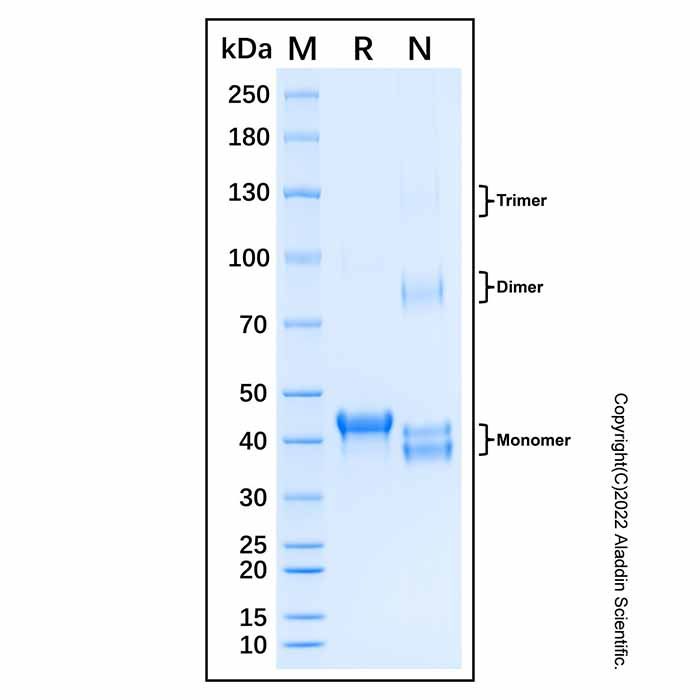
Recombinant Human SMAD2 Protein (rp220384) - SDS-PAGE 3 μg/lane of Recombinant Human SMAD2 Protein was resolved with SDS-PAGE under reducing (R) and non-reducing (N) conditions and visualized by Coomassie® Blue staining, showing a band at 43.3 kDa under reducing conditions and 38.4 & 41.8 & 82.4 & 126.2 kDa under non-reducing conditions. The conserved cysteine residues (Cys267, Cys330) on the surface of SMAD2 participate in disulfide bond formation. Under non-reducing conditions, these disulfide bonds remain intact, leading to distinct spatial conformations of the protein. Oligomerization is a critical step in SMAD2-mediated signal transduction (PMID: 10615055).

 ZJ25F0825944


 危险品化学品经营许可证(带存储)