
 K2222779
计算溶液所需的质量、体积或浓度。
为什么选择此级别
,适用于对基线干扰要求严格的色谱和分析工作流程。
储存与运输
-20°C储存,充氩。超低温运输 。请查阅批次 COA 获取详细规格。
质量文档
SDS、COA、产品数据表及规格说明书均可下载。可通过批号查询获取批次 COA。
文献证明
在色谱分析、有机合成和交叉偶联反应领域已被 16 篇同行评审文献引用。
概述
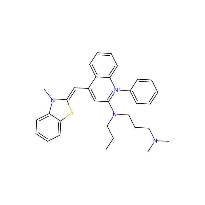
SYBR Green I是一种特制的不对称花菁染料,用于检测核酸。这种成分由N-烷基化苯并噻唑或苯并噁唑环系统组成,通过单次甲基桥连接吡啶鎓或喹啉鎓环系统。
核酸染料特点:
1. 相对安全:花菁染料,实验显示在凝胶染⾊浓度下⽆致突变性,可以代替致癌物溴化⼄锭EB作为各种核酸电泳的染⾊剂。
2. ⾼灵敏:紫外凝胶透射仪下灵敏度⾼ EB 染⾊法 5-10 倍,可⻅光透射仪下的灵敏度⽐ EB 染⾊法⾼20~30 倍。
3. 信噪⽐⾼:样品荧光信号强,⽆背景信号。
4. 操作简单:⽆须脱⾊或冲洗,即可⽤紫外凝胶透射仪观察或可⻅光透射仪观察。
5. 适⽤范围⼴:可适⽤于多种电泳分析,如琼脂糖凝胶电泳和 PAGE 凝胶电泳。
6. 使⽤⽅便:不影响其它修饰酶作⽤(如:Taq 酶、内切酶、T4 连接酶、反转录酶等)。
核酸染料使用方法:
1.胶染法(用法同EB)(推荐方法,见图1) 1) 制胶时加⼊ Sybr Green I 核酸染料。冷却胶到 50℃左右,每 100ml 胶中加⼊1-3µl Sybr Green I 核酸染料(⻅图 1)。
2) 按照常规⽅法进⾏电泳即可。
3) ⽤紫外凝胶透射仪或可⻅光透射仪观测。蓝光可透过玻璃, 观测聚丙烯酰胺凝胶时,可直接将托有凝胶的玻璃平⽫放⼊可⻅光透射仪内观测。 *注:此⽅法染⾊能准确确定⽚段分⼦量且⽤量较少。1ml 染料可以做 1000 块10 ml 胶,每块胶点 50 个样,可做 50000 次。
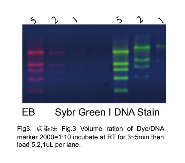
2.点染法(见图3)
1) 该⽅法适于琼脂糖凝胶电泳和PAGE凝胶电泳。
2) ⼯作液的配制:⽤电泳缓冲液将10000×的Sybr Green I 稀释100倍,即为Sybr Green I ⼯作液。Sybr Green I ⼯作液可以置2~8℃保存⼀个 ⽉以上, 浓缩液在-20℃保存半年。
3) 制胶:按常规⽅法制胶,不含任何染料。
4) 样品染⾊:向分析样品中加⼊Sybr Green I⼯作液和载样缓冲液,室温放置10分钟,使Sybr Green I与样品中DNA充分结合。Sybr Green I ⼯ 作液加⼊量为总上样量的1/5~1/10。
5) DNA Marker染⾊:将5μL DNA Marker、5μL DNA Marker稀释液和1μLSybr Green I ⼯作液混匀,室温放置5分钟,使Sybr Green I 与DNA 充分结合。
6) 上样、电泳:按常规操作。⽤紫外凝胶透射仪或可⻅光透射仪观测。蓝光可透过玻璃, 观测聚丙烯酰胺凝胶时,可直接将托有凝胶的玻璃平 ⽫放⼊可⻅光透射仪内观测。 *注:⽤点染法染⾊时,灵敏度最⾼,染料⽤量最少。通常点⼀个样加⼊ 1µL 即 可,可以使⽤ 10000 次, 但⼤⽚段稍有滞后现象,如果需要更准 确确定分⼦量(与 Marker 对⽐),建议使⽤胶染法。
3.泡染法
1) 按照常规⽅法进⾏制胶,其中不含任何染料。
2) ⽤ pH 7.0 - 8.5 的缓冲液(如:TAE, TBE),按照 1﹕1000 的⽐例稀释 Sybr Green I
3) 核酸染料,混匀,制成染⾊溶液。
4) 将染⾊溶液倒⼊合适的聚丙烯容器中,放⼊凝胶,⽤铝箔等盖住容器使染料避光。 室温振荡染⾊ 10-30 分钟,染⾊时间因凝胶浓度和厚度⽽ 定。聚丙烯酰胺凝胶直 接在玻璃平⽫上染⾊,将配好的稀释溶液轻轻地倒在胶板上,让稀释液均匀地覆 盖整个胶板,并染⾊ 30 分钟。玻璃 平⽫必须预先经过硅烷化溶液处理(避免染料 吸附在玻璃表⾯上)。
5) ⽤紫外凝胶透射仪或可⻅光透射仪观测。蓝光可透过玻璃, 观测聚丙烯酰胺凝胶时,可直接将托有凝胶的玻璃平⽫放⼊可⻅光透射仪内观测。 *注:⽤泡染⽅法染⾊时,可以精确确定核酸⽚段分⼦量。但染料⽤量是三种⽅法中⽤量最⼤的。 本品⽤ DMSO 溶解,因为 DMSO 溶点是 18.3℃,使⽤前请放置到室温充分溶解。
核酸染料使用注意事项:
1. Sybr Green核酸染料样品点染⽅法中,电泳不要超过 2 ⼩时,以免核酸染料从 DNA/RNA 上分离出来,产⽣弥散状条带。
2. ⽤点染⽅法染⾊时,条带稍有滞后现象,如果需要确定⽚段精确分⼦量(和 Marker 对⽐),建议⽤胶染法和泡染法。
3. 常规⽤酒精沉淀核酸过程中,Sybr Green I 核酸染料可以全部从核酸上去掉。
4. DNA电泳请选择 Sybr Green I 染料,RNA 电泳请选择 Sybr Green II染料,两种染料不通⽤。
5. Sybr Green I 核酸染料对玻璃和⾮聚丙烯材料具有⼀定亲合⼒。建议在稀释、贮存、染 ⾊等使⽤过程中⽤聚丙烯类容器。
6. 可以加Orange Red 作为标记. pH值在7.5-8.3之间,不要微波加热,加⼊热胶的温度低于50度。

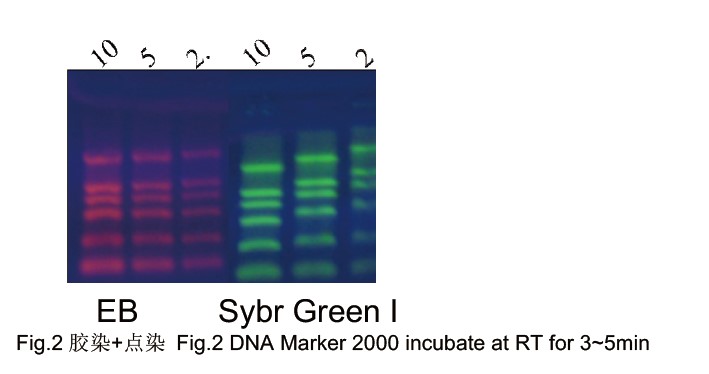

Sybr Green I and Sybr Green II Nucleic Acid Stains are produced by Dr.Chimin Du, are a kind of novel generation of fluorescent nucleic acid gel stains designed to replace the highly toxic ethidium bromide (EtBr). Sybr Green I is nontoxic and more sensitive than EtBr. Gels can be visualized under UV or Visible Light.
Features of Sybre Green Nucleic Acid Stains
1. Safety: Sybr Green is nontoxic and noncarcinogenic.
2. Ultra-sensitivity: It allows the visualization of as little as 20pg dsDNA, around 5-10 times more sensitive than EtBr under
UV and 8-20 times more sensitive than EtBr in Visible Light.
3. Convenience: NO need to rinse or wash gels. Add stain before load samples. Visualize gels under UV or Visible Light to avoid UV damage on DNA/RNA.
4. Wide range: suitable for agrose gel or PAGE.
5. No impact for the next experiments such as RT, PCR, enzyme digestion, and ligation.
6. Strong signal and no background
Directions for the Agrose Gels stained by Sybr Green
Protocol 1: Pre-cast Protocol (Add dye in the gel)
1.1 Prepare molten agarose gel solution using your standard protocol.
1.2 Add 1~3µl Sybr Green Nucleic Acid Stain per 50ml gel when the gel cool down to 50℃and mix thoroughly.
1.3 Cast the gel and allow it to solidify. Any leftover gel solution may be stored and reheated later for additional gel casting.
1.4 Load samples and run the gels using your standard protocol.
1.5 DNA stained with SYBR Green I stain can be readily visualized using a UV or blue-light sources (emit at 450, 473, 488, or 532 nm). Image the stained gel with the transilluminator and photograph the gel using.
Protocol 2: Post-staining Protocol (Stain Nucleic Acid after electrophoresis by adding dye in the gel stain solution)
2.1 Make gels: Do not add any nucleic acid stain when make gels.
2.2 Run gels as usual according to your standard protocol.
2.3 Prepare Sybr Green Nucleic Acid Staining solution: Dilute Sybr Green Nucleic Acid Stain with TAE or TBE (TBE (89 mM Tris base, 89 mM
boric acid, 1 mM EDTA, pH 8) and TAE (40 mM Trisacetate,1 mM EDTA, pH 8) on ratio 1:10000. Stain gels in the dark for 10-30min. Staining time depends on gel concentration and thickness. PAGE can be stained directly on the glass. Let staining solution cover PAGE gels for
30min. please use glassware to store staining solution or silicified glassware because stain will absorb on the glass.
2.4 Visualize gels a UV or blue-light sources.
2.5 Exact molecular weight can be measured by this method but the dyes are used much more in this way.
Protocol 3: Stain nucleic acid before electrophoresis (add dye in the loading buffer)
3.1. Prepare working solution: Dilute 10 µl Sybr Green Stain with 1ml running buffer TBE or TAE. This solution is stable up to one month at 4℃
3.2. Make gels: based on the routine method. Do not add any DNA/RNA stain in the gel.
3.3. Stain Nucleic Acid: Add 1µl Sybr Green I Nucleic Acid Stain working solution to 10µ mixture of sample and loading buffer, let it stay at RT for
3-5min for stain binding to nucleic acid completely. Normally, 1µL working solution is enough for one sample loading, and 1ml Sybr Green I Nucleic Acid Stain is enough to load 10,000 samples.
3.4. Stain markers: Mix 5µL Marker and 1µL Sybr Green I Nucleic Acid Stain working solution thoroughly, let it stay at RT for 5min to let Sybr
Green I Stain and DNA/RNA binding completely.
3.5. Load samples and run gels.
3.6. Visualize gels in UV or Visible Light to avoid UV damage on DNA/RNA.
*The big DNA fragments (>2Kb) will move slowly when bind to the stain. So please stain in DNA after electrophoresis or add stain in gels to measure molecular weight exactly.
Notes :
1. Do not run gels over 2 hrs. Or smeared bands appeared because Sybr Green I Stain will dissociate from DNA/RNA.
2. Sybr Green I Stain can dissociate from nucleic acids in ethanol.
3. Please stain nucleic acid in the gel or after electrophoresis to check the exact molecular weight of fragments when compared with molecular weight markers.
4. Please use EP tubes and other plastic wares in Sybr Green I Stain storage, dilution, and staining. Sybr Green I Stain can bind to glassware.
规格
英文别名
2-{[3-(dimethylamino)propyl](propyl)amino}-4-[(E)-(3-methyl-1,3-benzothiazol-2(3H)-ylidene)methyl]-1-phenylquinolinium | 2-{(E)-[2-{[3-(dimethylamino)propyl](propyl)amino}-1-phenylquinolin-4(1H)-ylidene]methyl}-3-methyl-1,3-benzothiazol-3-ium | N,N-dimeth
规格或纯度
10000 x in DMSO
英文名称
SYBR Green I Gel Staining Solution, 10000x
储存条件
-20°C储存,充氩
运输条件
超低温运输
名称和识别符
PubChem SID
EC号
838-836-0
分子类型
小分子
IUPAC Name
N,N-dimethyl-N'-[4-[(E)-(3-methyl-1,3-benzothiazol-2-ylidene)methyl]-1-phenylquinolin-1-ium-2-yl]-N'-propylpropane-1,3-diamine
INCHI
1S/C32H37N4S/c1-5-20-35(22-13-21-33(2)3)31-23-25(24-32-34(4)29-18-11-12-19-30(29)37-32)27-16-9-10-17-28(27)36(31)26-14-7-6-8-15-26/h6-12,14-19,23-24H,5,13,20-22H2,1-4H3/q+1
InChi Key
CGNLCCVKSWNSDG-UHFFFAOYSA-N
Smiles
CCCN(CCCN(C)C)C1=[N+](C2=CC=CC=C2C(=C1)C=C3N(C4=CC=CC=C4S3)C)C5=CC=CC=C5
Isomeric SMILES
CCCN(CCCN(C)C)C1=[N+](C2=CC=CC=C2C(=C1)/C=C/3\N(C4=CC=CC=C4S3)C)C5=CC=CC=C5
分子量
509.7
技术文档
高级数据
系统学分类
分类树(Taxonomy Tree)
| 界(kingdom) | 有机化合物 |
|---|---|
| 超类(Superclass) | 有机杂环化合物 |
| 类(Class) | 喹啉及其衍生物 |
| 亚类(Subclass) | 苯基喹啉 |
| 中间层级节点(Intermediate Tree Nodes) | 暂无 |
| 直接上位类(Direct Parent) | 苯基喹啉 |
| 其他上位类(Alternative Parents) | 氨基喹啉及其衍生物 苯并噻唑 二烷基芳胺 芳基硫醚 氨基吡啶及其衍生物 吡啶衍生物 苯及其取代衍生物 噻唑类 杂芳族化合物 三烷基胺 乙烯酮缩醛 氮杂环化合物 有机光子化合物 碳氢化合物衍生物 有机阳离子 |
| 分子骨架(Molecular Framework) | 芳香族杂多环化合物 |
| 取代基(Substituents) | 1,2-苯并噻唑 - 1,3-苯并噻唑 - 氨基酸 - 氨基吡啶 - 氨基吡啶 - 芳香族杂多环化合物 - 芳基硫醚 - 氮杂环 - 苯基化合物 - 苯并-噻唑 - 二烷基芳基胺 - 杂芳香族化合物 - 烃衍生物 - 酮烯缩醛或衍生物 - 单环苯基基团 - 有机铵盐 - 有机硝基化合物 - 有机氮化合物 - 有机氮化合物 - 苯基吡啶 - 吡啶 - 吡啶鎓 - 三元脂肪胺 - 三芳基胺 - 噻唑 |
| 描述(Description) | 该化合物属于苯基喹啉类有机化合物。这类杂环化合物含有被苯基取代的喹啉结构单元。 |
| 外部描述符(External Descriptors) | cyanine dye - tertiary amine - quinolines - benzothiazolium ion |
三维结构
交互式化学结构模型
化学和物理性质
敏感性
对光敏感;对湿度敏感
分子量
509.700 g/mol
XLogP3
8.200
氢键供体数Hydrogen Bond Donor Count
0
氢键受体数Hydrogen Bond Acceptor Count
4
可旋转键计数Rotatable Bond Count
9
精确质量Exact Mass
509.274 Da
单同位素质量Monoisotopic Mass
509.274 Da
拓扑极表面积Topological Polar Surface Area
38.900 Ų
重原子数Heavy Atom Count
37
形式电荷Formal Charge
1
复杂度Complexity
734.000
同位素原子数Isotope Atom Count
0
定义的原子立体中心计数Defined Atom Stereocenter Count
0
未定义的原子立体中心计数Undefined Atom Stereocenter Count
0
定义的键立体中心计数Defined Bond Stereocenter Count
1
未定义的键立体中心计数Undefined Bond Stereocenter Count
0
所有立体化学键的总数The total count of all stereochemical bonds
1
共价键合单元计数Covalently-Bonded Unit Count
1
安全和危险性(GHS)
象形图
信号词
警告
质检证书(CoA,COO,BSE/TSE 和分析图谱)
技术文档和文章
Understanding Cyanine Dyes: Structure & Mechanism, Channel Selection, Troubleshooting Essentials, and a Product Selection Roadmap (with 6 Category Tables)
目录: 技术文章
Fluorescent Probes: A Complete Beginner’s Guide — Definitions & Emission Mechanisms, Signal Readouts, Classification Framework, Selection Roadmap, and Product Tables (Tables 1–4)
目录: 技术文章
Role of ROX Passive Reference Dye in Real-Time Quantitative Fluorescence PCR: Functions and Practical Considerations
目录: 技术文章
Deoxyribonuclease (DNase): DNA Hydrolytic Cleavage Properties, Methodological Value, and Multi-Context Applications
目录: 技术文章
Agarose Precast Gels: Composition, Advantages, and Application Workflows for Standardized Nucleic Acid Electrophoresis Matrices
目录: 技术文章
Micrococcal Nuclease (MNase): A Technical Review of Enzymatic Properties, Reaction Control, and Research and Biomanufacturing Applications
目录: 技术文章
常用荧光素的比较与选型指南
目录: 技术文章
此产品的引用文献
溶液计算器
常见问题
本产品应如何储存?
请于?20 °C、氩气保护条件下保存。本品在氩气保护下供应,每次取用后请在惰性气体下重新密封。
本产品如何运输?
本产品采用保温箱配冰袋运输。收货后请立即拆包,并按上述储存条件保存。
本产品的 CAS 号、分子式和分子量是多少?
本产品的 CAS 号为 163795-75-3,分子式为 C32H37N4S,分子量为 509.7 g/mol。InChIKey CGNLCCVKSWNSDG-UHFFFAOYSA-N。
如何获取批次专用COA?
每个订单都会随附分析证书(COA),您也可以从账户订单历史中下载。如需订购前的COA,请联系您的客户代表,或使用PDP上的"申请COA"按钮——我们将提供代表性批次的COA。
产品仅提供给经过验证的企业、机构和合格的专业人员用于研发。不适用于人类、动物、诊断或治疗。
Copyright © 2023至今 上海阿拉丁生化科技股份有限公司 保留所有权利。


 危险品化学品经营许可证(带存储)