Rabbit anti Human AKAP 95 Antibody, Polyclonal, could be used for WB, IHC, IF/ICC and so on.
规格
产品名称
AKAP 95 Antibody
别名
蛋白激酶A锚定蛋白95抗体 | 蛋白激酶A锚定蛋白95多克隆抗体
英文别名
A kinase (PRKA) anchor protein 8 antibody | A kinase anchor protein 8 antibody | A kinase anchor protein 95 antibody | A kinase anchor protein 95 kDa antibody | A kinase anchor protein 95kDa antibody | A-kinase anchor protein 8 antibody | A-kinase anchor
规格或纯度
ExactAb™, 已验证, 无载体, 1.0 mg/mL
生化机理
Anchoring protein that mediates the subcellular compartmentation of cAMP-dependent protein kinase (PKA type II).
宿主种属
兔(Rabbit)
特异性
AKAP8
种属反应性
人(Human),大鼠(Rat),小鼠(Mouse)
免疫原
KLH-conjugated synthetic peptide encompassing a sequence within the center region of human AKAP 95.
阳性对照
WB: Hep G2, Jurkat and HEK293 cell lysates. IHC: Human heart tissue. IF/ICC: Jurkat cells.
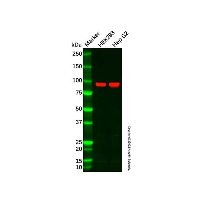
AKAP 95 Antibody (Ab087835) - Western Blot All lanes: AKAP 95 Antibody (Ab087835) at 1/1000 dilution Samples: Lysates at 20 µg per lane Secondary: Goat Anti-Rabbit IgG H&L (HRP) (Ab170144) at 1/40000 dilution Predicted band size: 76 kDa Observed band size: 91 kDa Exposure time: 60.0 s
AKAP 95 Antibody (Ab087835) - Western Blot All lanes: AKAP 95 Antibody (Ab087835) at 1/1000 dilution Samples: Lysates at 20 µg per lane Secondary: Goat Anti-Rabbit IgG H&L (HRP) (Ab170144) at 1/40000 dilution Predicted band size: 76 kDa Observed band size: 91 kDa Exposure time: 60.0 s
AKAP 95 Antibody (Ab087835) - Western Blot Immunohistochemical analysis of AKAP8 staining in human heart formalin fixed paraffin embedded tissue section. The section was pre-treated using heat mediated antigen retrieval with sodium citrate buffer (pH 6.0). The section was then incubated with AKAP 95 Antibody (Ab087835) at room temperature and detected using an HRP conjugated compact polymer system. DAB was used as the chromogen. The section was then counterstained with haematoxylin and mounted with DPX.
AKAP 95 Antibody (Ab087835) - IF/ICC Immunofluorescent analysis of AKAP8 staining in Jurkat cells. Formalin-fixed cells were permeabilized with 0.1% Triton X-100 in TBS for 5-10 minutes and blocked with 3% BSA-PBS for 30 minutes at room temperature. Cells were probed with AKAP 95 Antibody (Ab087835) in 3% BSA-PBS and incubated overnight at 4 °C in a humidified chamber. Cells were washed with PBST and incubated with a DyLight 594-conjugated secondary antibody (red) in PBS at room temperature in the dark.

 ZJ25F0117353


 危险品化学品经营许可证(带存储)