Rabbit anti Human GPCR GPR120 Antibody, Polyclonal, could be used for WB, IHC, ICC, IF and so on. Application: WB: 1/500-1/1000 IHC: 1/50-1/100
ICC/IF: 1/50-1/200 Protein Function: Receptor for medium and long-chain free fatty acid (FAA). Signals via a G(q)/G(11)-coupled pathway. Acts as a receptor for omega-3 fatty acids and mediates robust anti-inflammatory effects particulary in macrophages and fat cells. The anti-inflammatory effects involve inhibition of TAK1 through a beta-arrestin 2 (ARRB2)/TAB1 dependent effect but independent of G(q)/G(11)-coupled pathway. Mediates potent insulin sensitizing and antidiabetic effects by repressing macrophage-induced tissue inflammation. May mediates the taste of fatty acids.
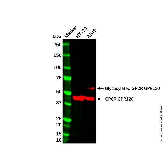
GPCR GPR120 Antibody (Ab106082) - Western Blot All lanes: GPCR GPR120 Antibody (Ab106082) at 1/1000 dilution Samples: Lysates at 20 µg per lane Secondary: Goat Anti-Rabbit IgG H&L (HRP) (Ab170144) at 1/10000 dilution Predicted band size: 40 kDa Observed band size: 57, 43 kDa Exposure time: 75.5 s
GPCR GPR120 Antibody (Ab106082) - IHC Immunohistochemical analysis of GPR120 staining in human prostate formalin fixed paraffin embedded tissue section. The section was pre-treated using heat mediated antigen retrieval with sodium citrate buffer (pH 6.0). The section was then incubated with GPCR GPR120 Antibody (Ab106082) at room temperature and detected using an HRP conjugated compact polymer system. DAB was used as the chromogen. The section was then counterstained with haematoxylin and mounted with DPX.
GPCR GPR120 Antibody (Ab106082) - IF/ICC Immunofluorescent analysis of GPR120 staining in LO2 cells. Formalin-fixed cells were permeabilized with 0.1% Triton X-100 in TBS for 5-10 minutes and blocked with 3% BSA-PBS for 30 minutes at room temperature. Cells were probed with GPCR GPR120 Antibody (Ab106082) in 3% BSA-PBS and incubated overnight at 4 °C in a hidified chamber. Cells were washed with PBST and incubated with a AF488-conjugated secondary antibody (green) in PBS at room temperature in the dark. Phalloidin-AF594 was used to stain Actin filaments (red). DAPI was used to stain the cell nuclei (blue).

 ZJ24F1113558


 危险品化学品经营许可证(带存储)