Rabbit anti Human HMGCS1 Antibody, Polyclonal, could be used for WB, IHC and so on. Application WB: 0.01-2µg/mL IHC: 5-20µg/mL IF/ICC: 2.5µg/mL Protein Function This enzyme condenses acetyl-CoA with acetoacetyl-CoA to form HMG-CoA, which is the substrate for HMG-CoA reductase.
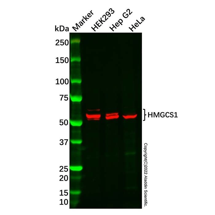
HMGCS1 Antibody (Ab108328) - Western Blot All lanes: HMGCS1 Antibody (Ab108328) at 1/1000 dilution Samples: Lysates at 20 µg per lane Secondary: Goat Anti-Rabbit IgG H&L (HRP) (Ab170144) at 1/20000 dilution
Predicted band size: 57 kDa Observed band size: 56, 57 kDa Exposure time: 43.4 s
HMGCS1 Antibody (Ab108328) - IHC DAB staining on IHC Samples: Human Liver Tissue Primary Ab: 10 µg/mL HMGCS1 Antibody (Ab108328) Second Ab: 2 µg/mL HRP-Linked Caprine Anti-Rabbit IgG Polyclonal Antibody.
HMGCS1 Antibody (Ab108328) - IHC DAB staining on IHC Samples: Human Brain Tissue Primary Ab: 10 µg/mL HMGCS1 Antibody (Ab108328) Second Ab: 2 µg/mL HRP-Linked Caprine Anti-Rabbit IgG Polyclonal Antibody.
HMGCS1 Antibody (Ab108328) - IHC DAB staining on IHC Samples: Human Kidney Tissue Primary Ab: 10 µg/mL HMGCS1 Antibody (Ab108328) Second Ab: 2 µg/mL HRP-Linked Caprine Anti-Rabbit IgG Polyclonal Antibody.
HMGCS1 Antibody (Ab108328) - IF/ICC IF analysis of HMGCS1 Antibody (Ab108328) in HeLa cells. The cells were fixed and permeabilized with 100% methanol for 5 minutes, and blocked with 5% BSA for 1 hour at room temperature. Cells were stained with HMGCS1 Antibody (Ab108328) at 1/200 dilution in blocking buffer overnight at 4°C, and then incubated with Goat Anti-Rabbit IgG H&L (AF647) (Ab181958) at a dilution of 1/1000 for 1 hour at room temperature in the dark (red). Cells were counterstained with DAPI (blue).

 ZJ24F0708247


 危险品化学品经营许可证(带存储)