Tyrosine-protein kinase that acts as a cell-surface receptor for the cytokine KITLG/SCF and plays an essential role in the regulation of cell survival and proliferation, hematopoiesis, stem cell maintenance, gametogenesis, mast cell development, migration and function, and in melanogenesis. In response to KITLG/SCF binding, KIT can activate several signaling pathways. Phosphorylates PIK3R1, PLCG1, SH2B2/APS and CBL. Activates the AKT1 signaling pathway by phosphorylation of PIK3R1, the regulatory subunit of phosphatidylinositol 3-kinase. Activated KIT also transmits signals via GRB2 and activation of RAS, RAF1 and the MAP kinases MAPK1/ERK2 and/or MAPK3/ERK1. Promotes activation of STAT family members STAT1, STAT3, STAT5A and STAT5B. Activation of PLCG1 leads to the production of the cellular signaling molecules diacylglycerol and inositol 1,4,5-trisphosphate. KIT signaling is modulated by protein phosphatases, and by rapid internalization and degradation of the receptor. Activated KIT promotes phosphorylation of the protein phosphatases PTPN6/SHP-1 and PTPRU, and of the transcription factors STAT1, STAT3, STAT5A and STAT5B. Promotes phosphorylation of PIK3R1, CBL, CRK (isoform Crk-II), LYN, MAPK1/ERK2 and/or MAPK3/ERK1, PLCG1, SRC and SHC1.
宿主种属
小鼠(Mouse)
特异性
Human c-Kit
种属反应性
人(Human)
免疫原
Recombinant protein corresponding to Human c-Kit
阳性对照
WB: K562, HeLa and Jurkat whole cell lysates IF/ICC: HEL cells
偶联
Unconjugated
克隆类型
单克隆抗体
Format
Whole IgG
轻链亚型
kappa
纯化方法
Protein A purified
浓度
1.0 mg/mL
分子类型
抗体
储存与运输
储存缓冲液
PBS, pH 7.2
储存条件
-20°C储存,避免反复冻融
运输条件
超低温运输
稳定性与储存
Store at 4°C short term (1-2 weeks). Store at -20°C or -80°C. Avoid freeze / thaw cycle.
图片
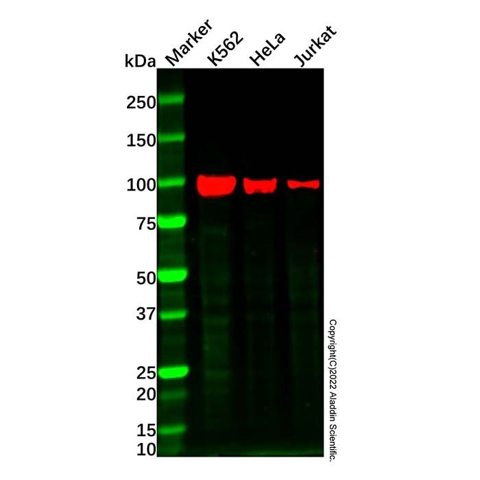
c-Kit Mouse mAb (Ab183891) - Western Blot All lanes: c-Kit Mouse Antibody (Ab183891) at 1/2000 dilution Samples: Lysates at 20 µg per lane Secondary: Goat Anti-Mouse IgG H&L (HRP) (Ab179001) at 1/20000 dilution
Predicted band size: 109 kDa Observed band size: 97 kDa Exposure time: 91.5 s
c-Kit Mouse mAb (Ab183891) - ICC/IF IF analysis of c-Kit (Green) in HEL cells. The cells were fixed and permeabilized with 100% methanol for 5 minutes, and blocked with 2% BSA for 1 hour at room temperature. Cells were stained with c-Kit Mouse mAb (Ab183891) at 5.0 μg/mL in blocking buffer for 2 hour at RT and then incubated with Goat Anti-Mouse IgG H&L (FITC) (Ab179003) at a dilution of 1/1000 for 1 hour at room temperature in the dark. Cells were counterstained with DAPI (Blue). Images were taken on the confocal laser scanning microscope.

 ZJ25F0622939

 危险品化学品经营许可证(带存储)