Rabbit anti Human ATP5L2 Antibody, Polyclonal, could be used for WB, IF/ICC and so on.
规格
产品名称
ATP5L2 Antibody
别名
ATP5L2抗体 | ATP5L2多克隆抗体
英文别名
ATP5K2 | ATP5L2 | ATP synthase subunit g 2, mitochondrial antibody | ATP synthase subunit g, mitochondrial antibody | ATP5K2 antibody | ATP5L antibody | ATP5L2 antibody | ATPase subunit g 2 antibody | ATPase subunit g antibody
规格或纯度
无载体, ExactAb™, 已验证, 1.0 mg/mL
生化机理
Mitochondrial membrane ATP synthase (F1F0 ATP synthase or Complex V) produces ATP from ADP in the presence of a proton gradient across the membrane which is generated by electron transport complexes of the respiratory chain. F-type ATPases consist of two structural domains, F1 - containing the extramembraneous catalytic core, and F0 - containing the membrane proton channel, linked together by a central stalk and a peripheral stalk. During catalysis, ATP synthesis in the catalytic domain of F1 is coupled via a rotary mechanism of the central stalk subunits to proton translocation. Part of the complex F0 domain. Minor subunit located with subunit a in the membrane (By similarity).
宿主种属
兔(Rabbit)
特异性
ATP5L2
种属反应性
人(Human),大鼠(Rat),小鼠(Mouse)
免疫原
A synthetic peptide derived from human ATP5L2
阳性对照
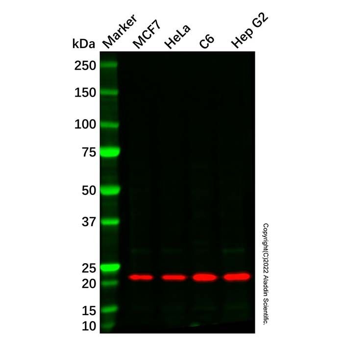
WB: Hep G2, HeLa, MCF7 and C6 cell lysates. IF/ICC: Hep G2 cells.
Store at 4°C short term (1-2 weeks). Store at -20°C long term (12 months). Upon receipt, it is recommended to aliquot. Avoid freeze/thaw cycle.
图片
ATP5L2 Antibody (Ab213100) - Western Blot All lanes: ATP5L2 Antibody (Ab213100) at 1/1000 dilution Samples: Lysates at 20 µg per lane Secondary: Goat Anti-Rabbit IgG H&L (HRP) (Ab170144) at 1/40000 dilution
Predicted band size: 11 kDa Observed band size: 21 kDa Exposure time: 16.1 s
ATP5L2 Antibody (Ab213100) - IF/ICC Immunofluorescent analysis of ATP5L2 staining in Hep G2 cells. Formalin-fixed cells were permeabilized with 0.1% Triton X-100 in TBS for 5-10 minutes and blocked with 3% BSA-PBS for 30 minutes at room temperature. Cells were probed with ATP5L2 Antibody (Ab213100) in 3% BSA-PBS and incubated overnight at 4 °C in a hidified chamber. Cells were washed with PBST and incubated with a DyLight 594-conjugated secondary antibody (red) in PBS at room temperature in the dark. DAPI was used to stain the cell nuclei (blue).

 ZJ25F0117127


 危险品化学品经营许可证(带存储)