半胱氨酸蛋白酶的Caspase家族已显示在凋亡中起关键作用。哺乳动物caspases可分为三个功能组:启动子半胱天冬酶(Caspase 2、8、9和10),执行者半胱天冬酶(Caspase-3、6和7)和炎性半胱天冬酶(Caspase1、4、5、11和12)。Caspase-8(也称为FLICE,MASH,Mch5)是半胱氨酸蛋白酶caspase家族的成员。在凋亡过程中,procaspase-8在天冬氨酸残基处通过自身蛋 白水解和/或被另一种caspase裂解而被加工。caspase-8加工后的活性形式由大和小的亚基组成,这些亚基缔合形成活性酶。活性caspase-8 已显示可激活caspase-3,从而导致细胞凋亡期间各种细胞靶蛋白的降解。
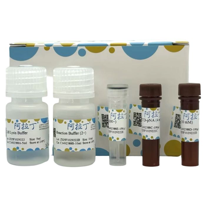
Caspase 8 活性检测试剂盒基于Caspase-8 水解肽底物Ac-IETD-pNA(acetyl-Ile-Glu-Thr-Asp p-nitroanilide),产生黄色的对硝基苯胺(pNA),pNA 在 405 nm 附近有强吸收,从而可以通过测定吸光度来检测Caspase-8的活性。
C1492388
| Component
| 20T | 50T
| 100T
| Storage
| C1492388A
| Cell Lysis Buffer
| 5 mL | 10 mL
| 20 mL
| 2-8℃
| C1492388B
| Reaction Buffer (2×)
| 10 mL
| 20 mL
| 40 mL
| 2-8℃
| C1492388C
| Ac-IETD-pNA (4 mM)
| 100 μL
| 250 μL
| 500 μL
| -20℃. Store in the dark.
| C1492388D
| pNA (10 mM)
| 100 μL
| 250 μL
| 500 μL
| -20℃. Store in the dark.
| C1492388E
| DTT (100×)
| 150 μL
| 400 μL
| 750 μL
| -20℃ |
|
自备耗材
酶标仪(能测405nm处的吸光度)
96孔板
低温离心机、制冰机
可调节式移液枪及枪头
去离子水、磷酸盐缓冲液(PBS)
匀浆器(如果是组织样本)
试剂准备
1、Working Cell Lysis Buffer:使用前,立即加入DTT(最终浓度:每1mL Cell Lysis Buffer加10µL DTT (100×)),置于冰上;4℃保存。
2、Working Reaction Buffer (1×):使用前,用去离子水稀释到Reaction Buffer (1×),再加入DTT(最终浓度:每1mL Reaction Buffer (1×)加 10 µL DTT (100×)),置于冰上;4℃保存。
3、Ac-IETD-pNA (4 mM):即用型;使用前,置于冰上;–20℃保存。用不完的试剂-20℃避光分装保存,避免反复冻融。
4、pNA (10 mM):即用型;使用前,置于冰上;–20℃避光保存。用不完的试剂-20℃避光分装保存,避免反复冻融。
5、DTT(100×):即用型;使用前,置于冰上;–20℃保存。用不完的试剂-20℃分装保存,避免反复冻融。
6、Standard 设置:按下表所示,用Reaction Buffer (1×,含DTT) 将标准品pNA (10mM) 稀释为200、100、50、25、12.5、0µM的标准溶液。
| 序号 | 标准品体积
| Reaction Buffer (1×, 含DTT) (µL)
| 标准品浓度 (μM)
| Std.1
| 6 µL pNA (10 mM)
| 294
| 200
| Std.2
| 150 µL of Std.1 (200 μmol/mL)
| 150 | 100
| Std.3
| 150 µL of Std.2 (100 μmol/mL)
| 150
| 50 | Std.4
| 150 µL of Std.3 (50 μmol/mL)
| 150
| 25 | Std.5
| 150 µL of Std.4 (25 μmol/mL)
| 150
| 12.5 | Std.6
| 0
| 150
| 0 |
|
样本制备
注意:我们建议使用新鲜样品。或按照“样本制备”将样品保存在-80℃。使用前,在冰上解冻,这可能会影响样品的稳定性,并且读数可能会低于预期。此试剂盒可检测蛋白水解活性。在样品制备步骤中请勿使用蛋白酶抑制剂,它可能会干扰测定。
1、通过所需方法诱导细胞凋亡。同时,建议对每个Caspase-8比色测定法设置不诱导的对照培养。
2、对于贴壁细胞,胰蛋白酶消化收集细胞。600g,4℃离心5min,小心吸去上清液。用1mL PBS洗涤细胞2次。离心后,去除上清液。在50µL Working Cell Lysis Buffer中重悬1-5×10⁶
个细胞。
3、对于悬浮细胞,600 g,4℃离心5 min,小心吸去上清液。用1 mL PBS洗涤细胞2次。离心后,去除上清液。在50µL Working Cell Lysis Buffer 中重悬 1-5×10
⁶
个细胞。
4、对于组织,将5-20 mg组织切成小块,用PBS清洗组织,加入0.1 mL Working Cell Lysis Buffer,冰浴匀浆。
5、裂解物冰上孵育15-20 min。
6、16,000 g,4℃离心15 min,然后将上清液转移至新试管中,置冰上待测。
7、立即测定Caspase-8的酶活性或-80℃保存样品。同时可以取少量样品用Bradford法测定蛋白浓度。
实验步骤
1、酶标仪预热30min以上,调节波长到405nm。
2、操作表(下述操作在96孔板中操作):
体积(μL)
| 空白孔
| 测定孔
| 对照孔
| 标准孔
| Working Reaction Buffer (1×,含 DTT)
| 100
| 50
| 55 | 0 | Standard
| 0 | 0 | 0 | 100 | 上清
| 0 | 50 | 50 | 0 | Ac-IETD-pNA (4 mM)
| 5 | 5 | 0 | 0 |
|
空白孔、测定孔、对照孔:
混匀,37℃孵育1-2h,检测405nm处吸光值。
空白孔记为A空,测定孔记为A测,对照孔记为A对。
计算ΔA测=A测-A空。
仅组织样本需要设置对照孔,对于组织样本,ΔA测=A测-A对。
标准孔:
立即检测405nm处吸光值,计算ΔA标=A标-AStd.6,AStd.6为不含pNA的标准孔的吸光值。
注意:
1、空白孔和标准孔只需测定 1-2 次。实验之前建议选择 2-3 个预期差异大的样本做预实验;
2、对于组织样本,样本本身有颜色,需要设置对照孔,细胞样本一般不需要设置对照孔;
3、在反应体系中,适当混匀,注意避免在混匀时产生气泡;
4、发现颜色变化比较明显时即可测定405nm的波长。如果颜色变化不明显,可以适当延长孵育时间至4h。
结果计算
注意:我们为您提供的计算公式,包括推导过程计算公式和简洁计算公式。两者完全相等。建议以加粗的简洁计算公式为最终计算公式。
1、按标准曲线计算
以标准溶液浓度为x轴,ΔA标为y轴,绘制标准曲线y=kx+b。将样本的ΔA测带入方程得到x值(μM)。
2、按酶活性增加百分比计算
Caspase-8 活性增加百分比=(实验处理组A测定-A空白)/(实验对照组A测定-A空白)×100%
该方法简单可靠,可粗略反应酶活性情况。
3. 按酶活计算
参考Chemicon公司的Caspase酶活力单位的定义:One unit is the amount of enzyme that will cleave 1.0 nmol of the colorimetric pNA-substrate per hour at 37℃ under saturated substrate concentrations。即一个酶活力单位定义为当底物饱和时,在 37℃一个小时内可以剪切1nmol pNA底物产生1nmol游离pNA的酶量。这样就可以计算出样品中含有多少个酶活力单位的Caspase酶活性。
Caspase-8 活性(U/mg prot)=x×V反总÷(V样×Cpr)÷T×10³=2.1×x÷Cpr÷T
V反总:反应体系总体积,0.105mL=1.05×10⁻⁴L;
V样:加入的样本体积,0.05mL;
T:反应时间,h;
Cpr:样本蛋白质浓度,mg/mL;
10³:单位换算系数,1μmol=10³nmol。
注意:对于样品中Caspase-8酶活力特别高的情况,须用Working Cell Lysis Buffer适当稀释样品后再进行测定。计算时再乘以稀释的稀释倍数n。
结果展示
经典标准曲线——以下数据和曲线仅供参考,实验者需根据自己的实验建立标准曲线。

The caspase family of cysteine proteases has been demonstrated to play a critical role in apoptosis. Mammalian caspases can be classified into three functional groups: initiator caspases (Caspase-2, -8, -9, and -10), executioner caspases (Caspase-3, -6, and -7), and inflammatory caspases (Caspase-1, -4, -5, -11, and -12). Caspase-8 (also known as FLICE, MACH, or Mch5) is a member of the caspase family of cysteine proteases. During apoptosis, procaspase-8 is processed through autoproteolysis and/or cleavage by other caspases at aspartic acid residues. The active form of processed caspase-8 consists of large and small subunits that associate to form the active enzyme. Active caspase-8 has been shown to activate caspase-3, leading to the degradation of various cellular target proteins during apoptosis.
The Caspase 8 Activity Assay Kit is based on the hydrolysis of the peptide substrate Ac-IETD-pNA (acetyl-Ile-Glu-Thr-Asp p-nitroanilide) by caspase-8, which releases yellow p-nitroaniline (pNA). pNA exhibits strong absorbance at approximately 405 nm, allowing the detection of caspase-8 activity through absorbance measurement.
C1492388
| Component
| 20T | 50T
| 100T
| Storage
| C1492388A
| Cell Lysis Buffer
| 5 mL | 10 mL
| 20 mL
| 2-8℃
| C1492388B
| Reaction Buffer (2×)
| 10 mL
| 20 mL
| 40 mL
| 2-8℃
| C1492388C
| Ac-IETD-pNA (4 mM)
| 100 μL
| 250 μL
| 500 μL
| -20℃. Store in the dark.
| C1492388D
| pNA (10 mM)
| 100 μL
| 250 μL
| 500 μL
| -20℃. Store in the dark.
| C1492388E
| DTT (100×)
| 150 μL
| 400 μL
| 750 μL
| -20℃ |
|
Materials Required but Not Provided
Microplate reader (capable of measuring absorbance at 405 nm)
96-well plate
Refrigerated centrifuge, ice maker
Adjustable pipettes and tips
Deionized water, phosphate-buffered saline (PBS)
Homogenizer (for tissue samples)
Reagent Preparation
Working Cell Lysis Buffer: Add DTT to Cell Lysis Buffer immediately before use (final concentration: 10 µL DTT (100×) per 1 mL Cell Lysis Buffer). Keep on ice; store at 4°C.
Working Reaction Buffer (1×): Dilute Reaction Buffer (2×) with deionized water to 1× concentration. Add DTT (final concentration: 10 µL DTT (100×) per 1 mL 1× Reaction Buffer). Keep on ice; store at 4°C.
Ac-IETD-pNA (4 mM): Ready-to-use. Thaw on ice before use; store at –20°C. Aliquot unused portions and avoid repeated freeze-thaw cycles.
pNA (10 mM): Ready-to-use. Thaw on ice before use; store at –20°C protected from light. Aliquot unused portions and avoid repeated freeze-thaw cycles.
DTT (100×): Ready-to-use. Thaw on ice before use; store at –20°C. Aliquot unused portions and avoid repeated freeze-thaw cycles.
Standard Curve Preparation: Prepare pNA standard solutions at 200, 100, 50, 25, 12.5, and 0 µM using Reaction Buffer (1×, containing DTT) according to the table below. Note: Prepare a fresh standard curve for each experiment. Diluted standards are unstable and must be used within 4 hours.
Standard
| Volume of Standard / Previous Standard
| Volume of Reaction Buffer (1×, with DTT) (µL)
| Concentration (μM)
| Std.1
| 6 µL pNA (10 mM)
| 294
| 200
| Std.2
| 150 µL of Std.1 (200 μmol/mL)
| 150 | 100
| Std.3
| 150 µL of Std.2 (100 μmol/mL)
| 150
| 50 | Std.4
| 150 µL of Std.3 (50 μmol/mL)
| 150
| 25 | Std.5
| 150 µL of Std.4 (25 μmol/mL)
| 150
| 12.5 | Std.6
| 0
| 150
| 0 |
|
Sample Preparation
Note: Fresh samples are recommended. Alternatively, store samples at -80°C after preparation. Thaw on ice before use, as freeze-thaw may affect stability and yield lower readings. Do not use protease inhibitors during sample preparation, as they may interfere with the assay.
Induce apoptosis in cells by an appropriate method. Include an uninduced control culture for each assay.
For adherent cells: Harvest by trypsinization. Centrifuge at 600 g, 4°C for 5 min. Carefully aspirate supernatant. Wash cell pellet twice with 1 mL PBS. Resuspend 1-5×10⁶ cells in 50 µL Working Cell Lysis Buffer.
For suspension cells: Centrifuge at 600 g, 4°C for 5 min. Carefully aspirate supernatant. Wash cell pellet twice with 1 mL PBS. Resuspend 1-5×10⁶ cells in 50 µL Working Cell Lysis Buffer.
For tissues: Mince 5-20 mg tissue into small pieces. Rinse with PBS. Homogenize in 0.1 mL Working Cell Lysis Buffer on ice.
Incubate lysates on ice for 15-20 min.
Centrifuge at 16,000 g, 4°C for 15 min. Transfer supernatant to a new tube and keep on ice.
Measure Caspase-8 activity immediately or store samples at -80°C. Determine protein concentration using a Bradford assay.
Assay Procedure
Preheat microplate reader for at least 30 min. Set wavelength to 405 nm.
Set up reactions in a 96-well plate as described below:
Volume (μL)
| Blank Well
| Test Well
| Control Well (For Tissue Samples)
| Standard Well
| Working Reaction Buffer (1×, with DTT)
| 100
| 50
| 55 | 0 | Standard
| 0 | 0 | 0 | 100 | Supernatant
| 0 | 50 | 50 | 0 | Ac-IETD-pNA(4 mM)
| 5 | 5 | 0 | 0 |
|
朱传侠For Blank, Test, and Control Wells:
Mix thoroughly and incubate at 37°C for 1–2 hours. Measure the absorbance at 405 nm.
Record the absorbance of the Blank Well as Ablank.
Record the absorbance of the Test Well as Atest.
Record the absorbance of the Control Well as Acontrol.
Calculate:
ΔAmeasured= Atest
–
Ablank
Note:
For tissue samples, use: ΔAmeasured= Atest – Acontrol.
For Standard Wells:
Measure the absorbance at 405 nm immediately after preparation.
Calculate:
ΔAstandard=Astandard-AStd.6
where AStd.6 is the absorbance of Standard Well 6 (0 μM pNA).
Notes:
Blank and Standard Wells require only 1-2 replicates.
Include a Control Well for tissue samples to account for inherent color.
Mix gently to avoid bubbles.
If color change is weak, extend incubation time up to 4 hours.
Calculations
Note: Provided formulas include a detailed derivation and a simplified version. They are equivalent. The simplified formula (in bold) is recommended for final calculation.
Standard Curve Method: Plot standard concentration (x-axis) against ΔAstandard(y-axis) to obtain the equation y = kx + b. Calculate the concentration (x, in µM) for the sample using its ΔAmeasured.
Percentage Increase in Activity:
Caspase-8
Activity Increase (%) = [(Atest_treated - Ablank / (Atest_control - Ablank)
] × 100%
Enzyme Activity Calculation (U/mg prot):
One unit of enzyme activity is defined as the amount of enzyme that cleaves 1.0 nmol of the colorimetric pNA substrate per hour at 37°C under saturated substrate concentrations (per Chemicon's definition).
Caspase-8
Activity (U/mg prot) = 2.1 × x ÷ Cpr÷ T
x: pNA concentration from standard curve (µM)
Cpr: Sample protein concentration (mg/mL)
T: Reaction time (hours)
2.1: Derived from (Vreaction_total× 10³) ÷ (Vsample) = (0.105 mL × 1000) ÷ (0.05 mL) = 2100, converted to 2.1 for the simplified formula.
Derivation details:
Caspase-8
Activity (U/mg prot) = (x × Vreaction_total) ÷ (Vsample × Cpr) ÷ T × 10³
Vreaction_total: Total reaction volume (0.105 mL = 1.05×10⁻⁴ L)
Vsample: Volume of sample added (0.05 mL)
10³: Conversion factor (1 µmol = 10³ nmol)
Note: For samples with very high Caspase-8 activity, dilute with Working Cell Lysis Buffer before assay and multiply the result by the dilution factor (n).朱传侠
Result Presentation
Representative Standard Curve: The following data and curve are for reference only. Users must generate their own standard curve for accurate quantification.

 ZJ25F1029222


 危险品化学品经营许可证(带存储)