
计算溶液所需的质量、体积或浓度。
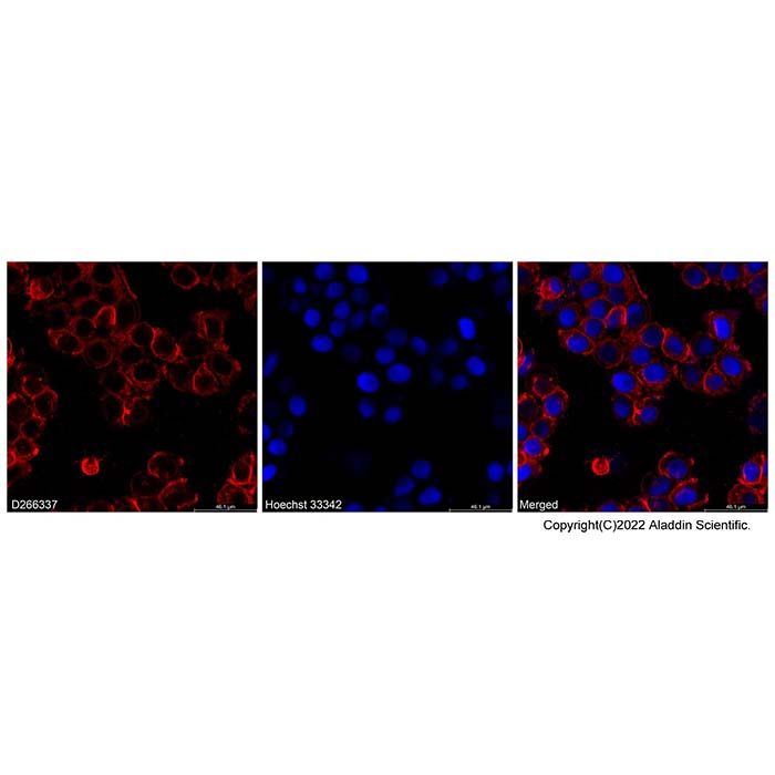
DiI iodide (D266337) - IF/ICC
IF analysis of DiI iodide (D266337) in HT-29 cells. Cells were incubated with 10 μM DiI at 37 °C for 20 minutes, followed by counterstaining with hoechst 33342 (B113845) (blue fluorescence).
BioReagent,生物染色剂,用于显微镜,适用于荧光分析 级 ,适用于对基线干扰要求严格的色谱和分析工作流程。
避光,-20°C储存。超低温运输 。请查阅批次 COA 获取详细规格。
SDS、COA、产品数据表及规格说明书均可下载。可通过批号查询获取批次 COA。
在色谱分析、有机合成和交叉偶联反应领域已被 0 篇同行评审文献引用。
DiD、DiO、DiI、DiR和DiS染料是一族亲脂性的荧光染料,可以用来染细胞膜和其它脂溶性生物结构。当与细胞膜结合后其荧光强度大大增强,这类染料有着很高的淬灭常数和激发态寿命。一旦对细胞染色,这类染料在整个细胞膜上扩散,最佳浓度时可以使整个细胞膜染色。它们的荧光颜色区分明显:DiI (橙色荧光),DiO (绿色荧光),DiD (红色荧光)和 DiR (深红色荧光)这使得他们可以用来对活细胞进行多色成像和流式分析。DiI和DiO可以分别用标准的 FITC和TRITC的滤光片。DiD可以用633 nm He-Ne激光器激发,有着比DiI更长的激发波长和发射波长,在细胞和组织染色中更有价值。 DiR的红外荧光可以穿透细胞和组织,在活体成像中用来示踪。
产品参数
Ex/Em (nm):549/565 (nm)
注意:Di系列染料是脂溶性染料,在缓冲溶液中容易析出,建议加入表面活性剂例如F127或者吐温20,终浓度千分之一。
使用方法
1. DiD,DiO,DiI,DiR和DiS细胞膜染色液制备
(1) 配置DMSO或EtOH储存液:储存液用DMSO或EtOH配置浓度1~5 mM。
注意:未使用的储存液保存在-20°C,避免反复冻融。
(2) 工作液制备:用合适的缓冲液 (如:无血清培养基,HBSS或PBS)稀释储存液,配制浓度为1~5 µM的工作液。
注意:工作液的最终浓度是根据不同细胞和实验的经验来配制。可以从推荐浓度的十倍以上寻找最佳条件。
2. 悬浮细胞染色
(1) 悬浮细胞密度为 1 × 10⁶/mL加入到工作液中。
(2) 在37℃培养细胞 2~20分钟,不同的细胞最佳培养时间不同。
(3) 染色细胞试管在1000~1500转离心5分钟。
(4) 倾倒上清液,再次缓慢加入预温37℃的培养液。
(5) 重复(3)、(4) 步骤两次以上。
3. 粘壁细胞的染色
(1) 使粘壁的细胞在无菌实验室培养。
(2) 从培养基中移走盖玻片,吸走过量培养液,将盖玻片放在潮湿的环境中。
(3) 在盖玻片的一角加入100μL的染料工作液,轻轻晃动使染料均匀覆盖所有细胞。
(4) 在37℃培养细胞 2~20分钟,不同的细胞最佳培养时间不同。
(5) 吸干染料工作液,用培养液洗盖玻片2~3次,每次用预温的培养基覆盖所有细胞,培养5~10分钟,然后吸干培养基。
4. 显微镜检测
(1) DiI滤光器选择XF32-Omega、31002-Chroma
(2) 多色染料的同时检测,滤光器按照以下设定:
a. DiI和DiO=Omega XF52,Chroma 51004;
b. DiI和DiD=Omega XF92,Chroma 51007;
c. DiI、DiO和DiD=Omega XF93,Chroma 61005
5. 流式细胞仪的检测
DiD、DiO、DiI、DiR和DiS染色的细胞可以分别用经典的FL1,FL2,FL3和FL4流式细胞仪检测。
DiD, DiO, DiI, DiR and DiS dyes are a family of lipophilic fluorescent dyes used for staining cell membranes and other lipid-soluble biological structures. Their fluorescence intensity is greatly enhanced upon membrane binding, and these dyes exhibit high quenching constants and long excited-state lifetimes. Once cells are stained, the dyes diffuse across the entire cell membrane, and at optimal concentrations, uniformly stain the whole membrane. They display distinct fluorescence colors: DiI (orange fluorescence), DiO (green fluorescence), DiD (red fluorescence) and DiR (deep-red fluorescence), enabling multicolor imaging and flow cytometry analysis of live cells. DiI and DiO can be used with standard FITC and TRITC filter sets, respectively. DiD is excitable by a 633 nm He-Ne laser and has longer excitation and emission wavelengths than DiI, making it more valuable for cell and tissue staining. The infrared fluorescence of DiR penetrates cells and tissues, allowing for in vivo imaging and tracing applications.
Product Parameters
Ex/Em (nm): 549/565 nm
Note: Di-series dyes are lipophilic and tend to precipitate in aqueous buffers. It is recommended to add a surfactant such as F127 or Tween-20 to a final concentration of 0.1%.
Protocol
1. Preparation of DiD, DiO, DiI, DiR and DiS Cell Membrane Staining Solutions
(1) Preparation of stock solutions in DMSO or EtOH:Prepare stock solutions at a concentration of 1-5 mM using DMSO or ethanol.
Note: Unused stock solutions should be stored at -20°C and avoid repeated freeze-thaw cycles.
(2) Preparation of working solutions:Dilute the stock solution with an appropriate buffer (e.g., serum-free medium, HBSS or PBS) to prepare a working solution at 1-5 µM.
Note: The final working concentration should be optimized based on specific cell types and experimental conditions. Optimal conditions may be determined starting from 10 times the recommended concentration.
2. Staining of Suspension Cells
(1) Add suspension cells at a density of 1 × 10⁶/mL to the working solution.
(2) Incubate the cells at 37°C for 2-20 minutes; optimal incubation time varies by cell type.
(3) Centrifuge the stained cell suspension at 1000-1500 rpm for 5 minutes.
(4) Discard the supernatant and gently add pre-warmed (37°C) culture medium.
(5) Repeat steps (3) and (4) at least twice.
3. Staining of Adherent Cells
(1) Culture adherent cells under sterile conditions.
(2) Remove coverslips from the culture medium, aspirate excess medium, and place coverslips in a humid environment.
(3) Add 100 μL of dye working solution to one corner of the coverslip and gently tilt to ensure uniform coverage of all cells.
(4) Incubate the cells at 37°C for 2-20 minutes; optimal incubation time varies by cell type.
(5) Aspirate the dye working solution and wash the coverslip 2-3 times with pre-warmed medium. For each wash, cover cells with medium, incubate for 5-10 minutes, then aspirate the medium completely.
4. Microscopy Detection
(1) Filter sets for DiI: XF32 (Omega), 31002 (Chroma)
(2) For simultaneous detection of multiple dyes, use the following filter sets:a. DiI & DiO: Omega XF52, Chroma 51004b. DiI & DiD: Omega XF92, Chroma 51007c. DiI, DiO & DiD: Omega XF93, Chroma 61005
5. Flow Cytometry Detection
Cells stained with DiD, DiO, DiI, DiR and DiS can be detected using conventional flow cytometers with FL1, FL2, FL3 and FL4 channels, respectively.
通过匹配包装上的批号来查找并下载产品的 COA,每批产品都进行了严格的验证,您可放心使用!
| 批号(Lot Number) | 证书类型 | 货号 |
|---|---|---|
 ZJ26F0535136 |
分析证书 | D266337 |
| ZJ26F0535137 |
分析证书 | D266337 |
我们的级别选择指南涵盖了目录中所有的变体的纯度、稳定剂状态和应用适用性。
查看 BioReagent 级别指南 → 查看 Biological Stain 级别指南 → 查看 for Fluorescence analysis 级别指南 → 查看 for Microscopy 级别指南 →

 危险品化学品经营许可证(带存储)