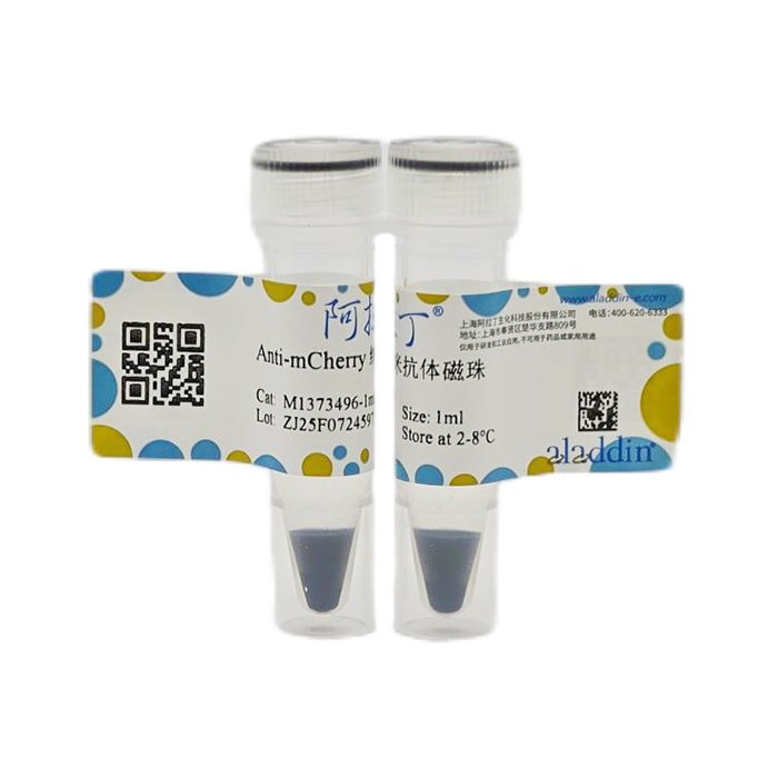
Anti-RFP 纳米抗体磁珠偶联了经过严格筛选、优化并重组表达的RFP纳米抗体,可用于捕获哺乳动物、植物、细菌、酵母、昆虫等多种生物细胞、组织提取物中的RFP融合蛋白及其相互作用蛋白。
实验前先将RFP蛋白与目标蛋白在细胞或组织中融合表达;之后将Anti-RFP 纳米抗体磁珠加入样本裂解液中,RFP纳米抗体与目标融合蛋白及其结合蛋白形成复合体;去除未结合的蛋白后,可以采用多种方法洗脱蛋白质,并且IP洗脱液中不会有抗体轻、重链污染。
产品特性:
磁珠直径
| 10-30 µm(磁性琼脂糖珠)
|
蛋白结合量
| ≥1.5 mg 蛋白/mL磁珠
|
反应性
| 可特异性结合各种常见类型的RFP蛋白(mRFP, mCherry, mRFPruby, mRuby2, tagRFP, mKate2, mPlum, mOrange, PA-mCherry, mScarlet),对融合蛋白N端、C端的RFP标签均可以识别。
|
应用
| 免疫沉淀(IP)、免疫共沉淀(Co-IP)、染色质免疫沉淀(CHIP)、RNA 结合蛋白免疫沉淀(RIP)
|
储存缓冲液
| 20 mM PBS, 5% BSA
|
储存条件
| 4℃. Do not freeze. |
产品优势:
1、纳米抗体,无轻重链污染。IP复合物无论采用非变性洗脱还是变性洗脱均不会出现抗体的轻、重链污染问题。
2、亲和力强。低nM级别亲和力,轻松应对低拷贝基因,或难转染细胞系。
3、结合量高。采用定向偶联技术每10 μL抗体磁珠可结合约15 μg重组蛋白。
4、特异性强。抗体磁珠通过了10株以上的空白细胞系检测不易产生非特异吸附。
5、兼容性好。无论标签在诱饵蛋白的N端或C端都可识别。
6、稳定性好。通过了40℃高温测试轻松应对物流失温,实验后忘记放冰箱等情况。
使用说明:
一、所需主要仪器
磁力架、混匀仪、低温离心机、超声破碎仪。
二、建议的缓冲液配方
缓冲液
| 配方 |
裂解缓冲液
| 10 mM Tris-HCl pH 7.5, 150 mM NaCl, 0.5 mM EDTA, 0.5% NP-40(在4°C下调整pH)
|
漂洗缓冲液
| 10 mM Tris-HCl pH 7.5, 150 mM NaCl, 0.5 mM EDTA, 0.05% NP-40(在4°C下调整pH)
|
洗脱缓冲液
| 200 mM 甘氨酸 pH 2.5
|
2×SDS-PAGE上样缓冲液
| 125 mM Tris-HCl pH 6.8, 4% SDS, 20%甘油, 0.004%溴酚蓝 |
三、注意事项
1、请勿干燥、冷冻或剧烈涡旋磁珠,禁止长时间置于磁场,可能会引起磁珠聚团,降低结合活性。
2、为保证磁珠均匀分布,请轻微涡旋或用枪头吹打混匀磁珠。
3、实验前应在裂解缓冲液和漂洗缓冲液中加入足量的蛋白酶抑制剂,RIP实验还应添加足量的RNase抑制剂,混合均匀,冰上保存,现配现用。
4、IP实验前,应先确认样本裂解液(input)中诱饵蛋白的表达水平。
5、每次IP实验应设置阴性对照组,通常采用表达RFP标签空载体的样本作为对照。
6、如果使用建议的缓冲体系不能获得很好的实验结果,可自行筛选、配制缓冲液进行实验。
7、具体样品量和孵育时间依赖于每个特定体系,可能需要优化才能得到最大产量。
8、当融合蛋白的分子量超过200kD时,磁珠IP效果可能受空间位阻影响而下降,需要测试用量。 9、为了您的安全和健康,请穿实验服并戴一次性手套操作。
10、本产品仅限于科学研究使用,不得用于临床诊断或治疗。
四、操作步骤
1、样本裂解(参考)
(1) 按如下方法收集样本:
样本类型
| 样本量/组
| 收集方法
|
动物细胞
| 1× 10⁷ 个
| 冷PBS漂洗2~3次,每次4℃ 500g离心5min收集沉淀,尽量吸干液体
|
动物组织
| 100~200 mg
| 冷PBS或生理盐水清洗干净,彻底去除血液等成分,液氮充分研磨
|
植物组织
| 200~300 mg
| 无菌双蒸水清洗干净,液氮充分研磨
|
革兰氏阴性细菌
| 50 μL 菌体沉淀
| 冷PBS漂洗2~3次,每次4℃ 5000 g离心5 min收集沉淀,尽量吸干液体
|
(2) 将样本置于冰上,每组样本加入500 μL预冷的裂解缓冲液,吹打混匀。
(3)
a. 动物细胞:最好冰上超声至溶液基本澄清;如果无超声条件,可以置于冰上裂解30min,间隔手动混匀。
b. 动物组织、植物组织、微生物:冰上超声破碎至溶液基本澄清。
(4) 4℃ 12000 g 离心 15 min,收集上清至新的离心管中,取30 μL作为input,剩余置于冰上备用或 -80℃保存。
【注意】:
i. 当样本不能完全裂解时(溶液很浑浊),可以增加裂解缓冲液、改善裂解方法或改善超声条件继续裂解,超声条件因样本类型和超声设备而异,应提前摸索好合适的条件。
ii. 裂解缓冲液用量可随样本量增加而等比例增加,但总孵育体积最大不超过离心管体积的2/3,体积过大可以更换大规格离心管。
2、免疫共沉淀
(1) 将Anti-RFP 纳米抗体磁珠上下颠倒混匀,每组取20 μL磁珠到新的离心管中。
(2) 加入200 µL漂洗缓冲液,颠倒混匀30次,放磁力架上静置1 min并弃上清。
(3) 重复上步操作一次。
(4) 向磁珠中加入相应组别的样本裂解液,放混匀仪上4℃孵育3 h至过夜。
(5) 放磁力架上静置1 min并弃上清。
(6) 加入500 μL漂洗缓冲液,颠倒混匀30次,放磁力架上静置1 min并弃上清。
(7) 重复上步操作两次,共漂洗三次。
3、洗脱
(1) SDS-PAGE 上样缓冲液洗脱(变性洗脱法):加入50 μL 1×SDS-PAGE上样缓冲液,95℃加热5~10min。放磁力架上静置1min或5000 g离心1min,收集上清至新的离心管中。洗脱后的蛋白 -20℃保存,或直接用于SDS-PAGE和Western Blot实验,SDS-PAGE胶条可以用于质谱检测。
(2) 甘氨酸洗脱(非变性洗脱法):加入40~50 µL 甘氨酸洗脱缓冲液,涡旋震荡20 s,放混匀仪上室温洗脱 10~15 min,涡旋震荡20 s;1000 g离心20 s,放磁力架上静置1 min,收集上清至新的离心管中。洗脱后的蛋白 -80℃保存,或直接用于SDS-PAGE、Western Blot和质谱等实验。
【备注:Anti-RFP 纳米抗体磁珠没有抗体轻重链污染问题,建议首选变性洗脱法,洗脱效率更高。】
Anti‑RFP Nanobody Magnetic Beads are coupled with a rigorously screened, optimized, and recombinantly expressed RFP nanobody. They can be used to capture RFP‑fusion proteins and their interacting proteins from cell or tissue extracts of various organisms including mammals, plants, bacteria, yeast, and insects.
Before the experiment, express the RFP protein fused to the target protein in cells or tissues. Then add Anti‑RFP Nanobody Magnetic Beads to the sample lysate; the RFP nanobody forms a complex with the target fusion protein and its binding partners. After removing unbound proteins, the proteins can be eluted by various methods, and the IP eluate will not be contaminated by antibody light or heavy chains.
Product Characteristics
Bead diameter
| 10–30 µm (magnetic agarose beads)
|
Protein binding capacity
| ≥1.5 mg protein / mL beads
|
Reactivity
| Specifically binds to various common types of RFP proteins (mRFP, mCherry, mRFPruby, mRuby2, tagRFP, mKate2, mPlum, mOrange, PA‑mCherry, mScarlet). Recognizes RFP tags at either the N‑ or C‑terminus of fusion proteins.
|
Applications
| Immunoprecipitation (IP), Co‑immunoprecipitation (Co‑IP), Chromatin Immunoprecipitation (ChIP), RNA‑Binding Protein Immunoprecipitation (RIP)
|
Storage buffer
| 20 mM PBS, 5% BSA
|
Storage conditions
| 4℃. Do not freeze. |
Product Advantages
1. Nanobody – no light/heavy chain contamination. Whether using native or denaturing elution, the IP complex will not show contamination from antibody light or heavy chains.
2. High affinity. Nanomolar‑level affinity allows easy handling of low‑copy‑number genes or difficult‑to‑transfect cell lines.
3. High binding capacity. Using oriented coupling technology, about 15 µg of recombinant protein can be bound per 10 µL of antibody‑coupled beads.
4. High specificity. The antibody‑coupled beads have been tested with over 10 blank cell lines and show minimal non‑specific adsorption.
5. Excellent compatibility. Recognizes tags at either the N‑ or C‑terminus of the bait protein.
6. Good stability. Passed 40°C high‑temperature testing, tolerates temporary temperature fluctuations during shipping or accidental omission of refrigeration after experiments.
Instructions for Use
1. Required Main Instruments
Magnetic rack, mixer/shaker, refrigerated centrifuge, ultrasonic disruptor.
2. Recommended Buffer Formulations
Buffer
| Formulation
|
Lysis Buffer
| 10 mM Tris‑HCl pH 7.5, 150 mM NaCl, 0.5 mM EDTA, 0.5% NP‑40 (adjust pH at 4°C)
|
Wash Buffer
| 10 mM Tris‑HCl pH 7.5, 150 mM NaCl, 0.5 mM EDTA, 0.05% NP‑40 (adjust pH at 4°C)
|
Elution Buffer
| 200 mM glycine pH 2.5
|
2× SDS‑PAGE Loading Buffer
| 125 mM Tris‑HCl pH 6.8, 4% SDS, 20% glycerol, 0.004% bromophenol blue |
3. Precautions
3.1 Do not dry, freeze, or vortex the beads vigorously. Avoid prolonged placement on a magnetic rack, as this may cause bead aggregation and reduce binding activity.
3.2 To ensure uniform bead distribution, gently vortex or mix by pipetting up and down.
3.3 Before the experiment, add sufficient protease inhibitors to the lysis and wash buffers. For RIP experiments, also add sufficient RNase inhibitors. Mix well and keep on ice; prepare freshly.
3.4 Before IP experiments, confirm the expression level of the bait protein in the sample lysate (input).
3.5 Each IP experiment should include a negative control group, typically using a sample expressing the RFP tag empty vector.
3.6 If the recommended buffer system does not yield satisfactory results, you may screen and prepare your own buffers.
3.7 The specific sample amount and incubation time depend on each particular system and may require optimization for maximum yield.
3.8 When the molecular weight of the fusion protein exceeds 200 kDa, bead‑based IP efficiency may decrease due to steric hindrance, and the bead amount may need to be tested.
3.9 For your safety and health, please wear a lab coat and disposable gloves during operation.
3.10 This product is for scientific research only and must not be used for clinical diagnosis or treatment.
4. Operating Steps
4.1 Sample Lysis (Reference)
(1) Collect samples as follows:
Sample Type
| Amount per Group
| Collection Method
|
Animal cells
| 1 × 10⁷ cells
| Wash 2–3 times with cold PBS, centrifuge at 500 g, 4°C for 5 min each time to collect the pellet; remove liquid as completely as possible.
|
Animal tissue
| 100~200 mg
| Rinse thoroughly with cold PBS or saline to remove blood, etc.; grind thoroughly under liquid nitrogen.
|
Plant tissue
| 200~300 mg
| Wash with sterile double‑distilled water; grind thoroughly under liquid nitrogen.
|
Gram‑negative bacteria
| 50 µL bacterial pellet
| Wash 2–3 times with cold PBS, centrifuge at 5000 g, 4°C for 5 min each time to collect the pellet; remove liquid as completely as possible.
|
(2) Place samples on ice. Add 500 µL of pre‑chilled lysis buffer per group and mix by pipetting.
(3)
a. Animal cells: Preferably sonicate on ice until the solution is essentially clear. If no sonication is available, lyse on ice for 30 min with occasional manual mixing.
b. Animal tissue, plant tissue, microorganisms: Sonicate on ice until the solution is essentially clear.
(4) Centrifuge at 4°C, 12,000 g for 15 min. Transfer the supernatant to a new tube. Take 30 µL as “input”; keep the remainder on ice or store at –80°C.
Notes:
i. If the sample is not completely lysed (solution very turbid), increase the lysis buffer volume, improve the lysis method, or adjust sonication conditions. Optimal sonication conditions vary by sample type and equipment and should be established beforehand.
ii. The lysis buffer volume can be increased proportionally with sample amount, but the total incubation volume should not exceed 2/3 of the tube capacity. For larger volumes, use a larger tube.
4.2 Co‑immunoprecipitation
(1) Gently invert the Anti‑RFP Nanobody Magnetic Beads to mix. Transfer 20 µL of beads per group to a new tube.
(2) Add 200 µL of wash buffer, invert to mix 30 times, place on a magnetic rack for 1 min, and discard the supernatant.
(3) Repeat the previous step once.
(4) Add the corresponding group’s sample lysate to the beads. Incubate on a shaker at 4°C for 3 h to overnight.
(5) Place on a magnetic rack for 1 min and discard the supernatant.
(6) Add 500 µL of wash buffer, invert to mix 30 times, place on a magnetic rack for 1 min, and discard the supernatant.
(7) Repeat the previous step twice, for a total of three washes.
4.3 Elution
(1) SDS‑PAGE loading buffer elution (denaturing elution): Add 50 µL of 1× SDS‑PAGE loading buffer and heat at 95°C for 5–10 min. Place on a magnetic rack for 1 min or centrifuge at 5000 g for 1 min, then transfer the supernatant to a new tube. The eluted proteins can be stored at –20°C or used directly for SDS‑PAGE and Western blotting. SDS‑PAGE gel bands can be used for mass spectrometry.
(2) Glycine elution (native elution): Add 40–50 µL of glycine elution buffer, vortex for 20 s, incubate on a shaker at room temperature for 10–15 min, vortex again for 20 s. Centrifuge at 1000 g for 20 s, place on a magnetic rack for 1 min, and transfer the supernatant to a new tube. The eluted proteins can be stored at –80°C or used directly for SDS‑PAGE, Western blotting, mass spectrometry, etc.
Note: Anti‑RFP Nanobody Magnetic Beads do not suffer from antibody light/heavy chain contamination. Denaturing elution is recommended first, as it provides higher elution efficiency.

 ZJ26F1039800


 危险品化学品经营许可证(带存储)