
计算溶液所需的质量、体积或浓度。
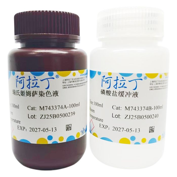
瑞氏-姬姆萨染色试剂盒
有货
BioReagent,生物染色剂,用于显微镜 级 ,适用于对基线干扰要求严格的色谱和分析工作流程。
避光,室温。常规运输 。请查阅批次 COA 获取详细规格。
SDS、COA、产品数据表及规格说明书均可下载。可通过批号查询获取批次 COA。
在色谱分析、有机合成和交叉偶联反应领域已被 0 篇同行评审文献引用。
瑞氏色素是酸性染料伊红 (Eosin) 和碱性染料亚甲蓝 (Methylene Blue) 组成的复合染料,对原生质的染色有很好的区别作用。姬姆萨染液由天青Ⅱ与伊红混合而成,染色原理和结果与瑞氏染色法基本相同,姬姆萨染色液对胞浆着色力较强,能较好的显示胞浆的嗜碱性程度,特别是对血液和骨髓细胞中的嗜天青、嗜酸性、嗜碱性颗粒,着色清晰,但是对胞核着色偏深,核结构显色不佳,故姬姆萨染液常和瑞氏染液联合使用。
瑞氏-姬姆萨复合染色试剂盒以优质瑞氏色素和姬姆萨色素为主要原料,能呈现出清晰的细胞染色效果。经常用于血液和细胞涂片、骨髓细胞涂片、细菌染色。细胞质呈红色,细胞核及细菌呈蓝色,嗜酸性颗粒呈橘红色。染液中加中性甘油,防止甲醇挥发或氧化,同时也可使血细胞染色较清晰。该染液的特点:由瑞氏-姬姆萨复合染色液和磷酸盐缓冲液组成,等量混合使用或分别处理标本使用。
| M743374 | Component | 2×100 mL | 2×500 mL | Storage |
| M743374A | 瑞氏姬姆萨染色液 | 100 mL | 500 mL | RT. Store in the dark |
| M743374B | 磷酸盐缓冲液 | 100 mL | 500 mL | RT. |
使用说明:
1、常规方法制备血液涂片或骨髓涂片或细菌涂片,待涂片自然干燥。
2、将血液涂片或骨髓涂片置于染色架上。
3、滴加适量瑞氏姬姆萨染色液覆盖涂片,室温染色1-2min。
4、涂片滴加等量磷酸盐缓冲液,轻轻晃动玻片或采用其他方式混合,使磷酸盐缓冲液与瑞氏姬姆萨染色液混匀,室温静置3-10min。
5、步骤 3、4 亦可以采用如下方法:取瑞氏姬姆萨染色液和磷酸盐缓冲液等量混合,即为瑞氏姬姆萨工作液,滴加该工作液于血液涂片或骨髓涂片上,室温静置3-10min。
6、用自来水或蒸馏水从玻片一端轻轻冲洗。 (注:也可用磷酸盐缓冲液等量稀释后,冲洗玻片,时间控制在30s左右)
7、干燥。镜检:先用低倍镜观察血涂片,再用油镜。
注意事项:
1、血液涂片或骨髓涂片应厚薄均匀,以免影响染色效果。
2、涂片染色中,请勿先去除染液或直接对涂片用力冲洗。不能先倒掉染液,以免染料沉着于涂片上。
3、染色液可重复使用,但不能多次重复,若有沉淀物应过滤后使用。
4、染色过深可用甲醇或乙醇适当脱色,最好不复染。
5、如果染色过深或过浅,应调整染色时间或工作液浓度。
6、为了您的安全和健康,请穿实验服并戴一次性手套操作。
染色结果:
| 细菌、细胞核 | 蓝色 |
| 组织细胞的细胞质、血红蛋白、嗜酸性颗粒 | 粉红或橘红色 |
Wright stain is a compound dye composed of the acidic dye Eosin and the basic dye Methylene Blue, which delivers excellent differential staining of protoplasm. Giemsa stain is a mixture of Azure II and Eosin. Its staining principle and resultant coloration are basically consistent with Wright staining. Giemsa stain exhibits strong affinity for cytoplasm and clearly reveals the basophilic degree of cytoplasm. In particular, it distinctly stains azurophilic, eosinophilic and basophilic granules in blood and bone marrow cells. However, it overstains cell nuclei and fails to display fine nuclear structures clearly. For this reason, Giemsa stain is frequently used in combination with Wright stain.
Wright-Giemsa Staining Kit is formulated with high-quality Wright and Giemsa dyes as the primary raw materials, yielding sharp and clear cellular staining results. It is widely applied for staining blood smears, cell smears, bone marrow smears and bacteria. Cytoplasm stains red; cell nuclei and bacteria appear blue; eosinophilic granules turn orange-red. Neutral glycerol is added to the staining solution to inhibit methanol volatilization and oxidation, while also sharpening the staining morphology of blood cells. Features of this stain: The kit consists of Wright-Giemsa combined staining solution and phosphate buffer. The two components can be mixed in equal volumes for use, or applied to specimens separately.
| M743374 | Component | 2×100 mL | 2×500 mL | Storage |
| M743374A | Wright-Giemsa Staining Solution | 100 mL | 500 mL | RT. Store in the dark |
| M743374B | Phosphate Buffer Solution | 100 mL | 500 mL | RT. |
Instructions for Use:
1. Prepare blood smears, bone marrow smears or bacterial smears via standard protocols and allow the smears to air-dry naturally.
2. Place the blood or bone marrow smears on a staining rack.
3. Add an appropriate amount of Wright-Giemsa staining solution to fully cover the smear, and stain at room temperature for 1–2 minutes.
4. Add an equal volume of phosphate buffer solution to the smear. Gently rock the slide or mix by other means to fully blend the buffer with the Wright-Giemsa staining solution, then incubate at room temperature for 3–10 minutes.
5. Alternative procedure for Steps 3 and 4: Mix equal volumes of Wright-Giemsa staining solution and phosphate buffer to prepare Wright-Giemsa working solution. Apply the working solution to blood or bone marrow smears and incubate at room temperature for 3–10 minutes.
6. Gently rinse the slide from one end with tap water or distilled water. (Note: The slide can also be rinsed with phosphate buffer diluted at an equal ratio for approximately 30 seconds.)
7. Air-dry the slide. Microscopic examination: Observe the blood smear under low magnification first, then switch to oil immersion objective.
Precautions:
1. Blood and bone marrow smears must be uniformly thick to avoid uneven staining results.
2. Do not pour off the staining solution in advance or flush the smear with strong water flow during staining. Premature draining of the stain will cause dye precipitate to deposit on the smear.
3. The staining solution may be reused a limited number of times. Filter the solution to remove precipitates before reusing.
4. Over-stained slides can be appropriately decolorized with methanol or ethanol; re-staining is not recommended.
5. Adjust the staining duration or working solution concentration if staining appears too dark or too pale.
6. Wear a lab coat and disposable gloves during all operations for personal safety and health protection.
Staining Results:
| Bacteria, Cell nuclei | Blue |
| Cytoplasm of tissue cells, hemoglobin, eosinophilic granules | Pink or orange-re |
通过匹配包装上的批号来查找并下载产品的 COA,每批产品都进行了严格的验证,您可放心使用!
| 批号(Lot Number) | 证书类型 | 货号 |
|---|---|---|
 ZJ26F0838129 ZJ26F0838129 |
分析证书 | M743374 |
| ZJ26F0838128 |
分析证书 | M743374 |
| ZJ26F0131662 |
分析证书 | M743374 |
| ZJ25F0724232 |
分析证书 | M743374 |
| ZJ25F0521433 |
分析证书 | M743374 |
我们的级别选择指南涵盖了目录中所有的变体的纯度、稳定剂状态和应用适用性。
查看 BioReagent 级别指南 → 查看 Biological Stain 级别指南 → 查看 for Microscopy 级别指南 →

 危险品化学品经营许可证(带存储)