ERBB4 is a single-pass type I membrane protein with multiple cysteine rich domains, a transmembrane domain, a tyrosine kinase domain, a phosphotidylinositol-3 kinase binding site and a PDZ domain binding motif. ERBB4 is expressed at highest levels in brain, heart, kidney, in addition to skeletal muscle, parathyroid, cerebellum, pituitary, spleen, testis and breast. And lower levels in thymus, lung, salivary gland, and pancreas. It specifically binds to and is activated by neuregulins, NRG-2, NRG-3, heparin-binding EGF-like growth factor, betacellulin and NTAK. ERBB4 also can be activated by other factors and induces a variety of cellular responses including mitogenesis and differentiation. ERBB4 regulates development of the heart, the central nervous system and the mammary gland, gene transcription, cell proliferation, differentiation, migration and apoptosis. It is required for normal cardiac muscle differentiation during embryonic development, and for postnatal cardiomyocyte proliferation. ERBB4 also play a role on the normal development of the embryonic central nervous system, especially for normal neural crest cell migration and normal axon guidance. It is required for mammary gland differentiation, induction of milk proteins and lactation.
Post-translational: Isoform JM-A CYT-1 and isoform JM-A CYT-2 are processed by ADAM17. Proteolytic processing in response to ligand or 12-O-tetradecanoylphorbol-13-acetate stimulation results in the production of 120 kDa soluble receptor forms and intermediate membrane-anchored 80 kDa fragments (m80HER4), which are further processed by a presenilin-dependent gamma-secretase to release a cytoplasmic intracellular domain (E4ICD; E4ICD1/s80Cyt1 or E4ICD2/s80Cyt2, depending on the isoform). Membrane-anchored 80 kDa fragments of the processed isoform JM-A CYT-1 are more readily degraded by the proteasome than fragments of isoform JM-A CYT-2, suggesting a prevalence of E4ICD2 over E4ICD1. Isoform JM-B CYT-1 and isoform JM-B CYT-2 lack the ADAM17 cleavage site and are not processed by ADAM17, precluding further processing by gamma-secretase. Autophosphorylated on tyrosine residues in response to ligand binding. Autophosphorylation occurs in trans, i.e. one subunit of the dimeric receptor phosphorylates tyrosine residues on the other subunit. Ligands trigger phosphorylation at specific tyrosine residues, thereby creating binding sites for scaffold proteins and effectors. Constitutively phosphorylated at a basal level when overexpressed in heterologous systems; ligand binding leads to increased phosphorylation. Phosphorylation at Tyr-1035 is important for interaction with STAT1. Phosphorylation at Tyr-1056 is important for interaction with PIK3R1. Phosphorylation at Tyr-1242 is important for interaction with SHC1. Phosphorylation at Tyr-1188 may also contribute to the interaction with SHC1. Isoform JM-A CYT-2 is constitutively phosphorylated on tyrosine residues in a ligand-independent manner. E4ICD2 but not E4ICD1 is phosphorylated on tyrosine residues. Ubiquitinated. During mitosis, the ERBB4 intracellular domain is ubiquitinated by the APC/C complex and targeted to proteasomal degradation. Isoform JM-A CYT-1 and isoform JM-B CYT-1 are ubiquitinated by WWP1. The ERBB4 intracellular domain (E4ICD1) is ubiquitinated, and this involves NEDD4.
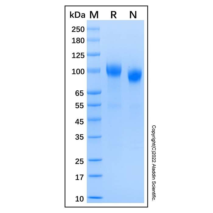
87.5-109.6 kDa, under reducing conditions; 78.7-104.3 kDa, under non-reducing conditions.
分子类型
蛋白质
产品线
生物产品
储存与运输
物理形态
冻干(Lyophilized)
储存缓冲液
Lyophilized from PBS, pH 7.4.
浓度
见COA
复溶
We recommend that this vial be briefly centrifuged prior to opening to bring the contents to the bottom. Reconstitute at 1.0 mg/mL in sterile distilled water. Stock solutions should be apportioned into working aliquots and stored at ≤ -20 °C. Further dilutions should be made in appropriate buffered solutions.
储存条件
-20°C储存,避免反复冻融
运输条件
超低温运输
稳定性与储存
Store at -20°C for 1 year. Avoid freeze/thaw cycle.
图片
Recombinant Human ErbB4/HER4 Protein (rp169609) - SDS-PAGE 3 μg/lane of Recombinant Human ErbB4/HER4 Protein was resolved with SDS-PAGE under reducing (R) and non-reducing (N) conditions and visualized by Coomassie® Blue staining, showing a band at 87.5-109.6 kDa under reducing conditions and 78.7-104.3 kDa under non-reducing conditions.

 ZJ25F0420760


 危险品化学品经营许可证(带存储)