一步法 LumiDye™ 555 鬼笔环肽/核蓝双染试剂盒是一款可同时实现F-肌动蛋白与细胞核特异性双标染色的即用型试剂盒,核心组分为LumiDye™Phalloidin与Nucleiblue,专为解决细胞骨架与细胞核共定位分析的实验需求设计。本产品的核心原理为:
(1) LumiDye™Phalloidin作为鬼笔环肽衍生物,可与真核生物细胞内的丝状肌动蛋白(F-actin)发生高亲和力特异性结合,且不与单体G-肌动蛋白结合,能精准标记细胞骨架微丝结构。
(2) Nucleiblue作为高特异性核荧光染料,可与细胞核DNA特异性结合,实现细胞核的清晰荧光标记,二者协同作用可完成F-肌动蛋白与细胞核的同步染色。
相较于传统的分开配制型肌动蛋白与核染色试剂,本产品为即用型预混体系,无需实验人员自行稀释、配比,大幅节省实验操作时间,且试剂配比经过优化,非特异性染色可忽略不计;同时LumiDye™Phalloidin分子量小,与F-actin结合后不影响肌动蛋白结合蛋白的相互作用,进一步保留F-actin生物学功能;LumiDye™染料是我司开发的一系列新一代荧光染料,与其他荧光染料相比,在亮度,光稳定性和水溶性方面具有综合优势。其中LumiDye™Phalloidin可稳定F-actin结构,阻止其解聚,保证微丝形态的完整呈现,且对不同物种的F-actin结合亲和力无显著差异,基本不影响F-actin的功能,标记后的肌动蛋白丝仍保持正常功能,标记的肌纤维能够收缩,标记的肌球蛋白可继续移动,同时在各类植物、动物细胞中,对大、小微丝具有相似的亲和力,以1∶1的化学计量比与肌动蛋白亚基结合,可直接用于细胞中F-actin的定量研究;Nucleiblue荧光信号亮度高、光稳定性强,背景干扰极低,可清晰显示细胞核的形态、位置及数量变化。多重优势让定量与定性分析均可高效实现。本产品可广泛应用于荧光显微镜(含激光共聚焦显微镜)观察、流式细胞术检测,同时适用于细胞生物学基础研究、细胞骨架动态变化分析等实验场景。
肌动蛋白作为细胞骨架的核心组成部分,其丝状形式F-actin的形态、分布与细胞核的结构变化密切关联,是反映细胞迁移及形态重塑的重要指标,而鬼笔环肽因对F-actin的特异性结合特性,成为肌动蛋白染色的经典工具,Nucleiblue则是新一代高特异性细胞核荧光染色试剂,凭借优异的染色性能成为细胞核标记的优选工具,二者联用是细胞形态与亚细胞结构分析的核心方法。
本试剂盒在该科学背景下,将LumiDye™Phalloidin与Nucleiblue进行一体化优化配制,且LumiDye™Phalloidin兼具标记精准、功能保留与定量适配的特性,在荧光显微镜观察中可清晰呈现F-肌动蛋白的微丝分布与细胞核的位置关系,实现亚细胞结构的精准共定位分析;在流式细胞术中可同时对F-actin表达水平进行精准定量检测;在药物筛选研究中,可快速检测药物处理后细胞骨架的重塑与细胞核的形态改变,为药物的作用机制研究提供直观的形态学依据;在细胞生物学基础研究中,可用于分析细胞迁移、侵袭、分化过程中F-actin与细胞核的动态变化,且能在保留F-actin生理功能的前提下开展相关研究,最大程度还原细胞真实生理状态。本产品以高特异性、高稳定性、高便捷性与强功能性为核心优势,为实验人员提供了高效、准确的细胞骨架-细胞核双标分析工具。
应用范围:细胞、冷冻组织切片、酵母及真菌的F-actin的标记、免疫荧光兼容
产品特点:
⁕ 双标一体,即用型配方:预混LumiDye™Phalloidin与Nucleiblue染色试剂,开盖即用,无需稀释、混合与滴定,一步完成F-actin 与细胞核染色,大幅简化实验流程。
⁕ 信号精准,共定位准:LumiDye™Phalloidin高特异性结合F-actin,不识别G‑肌动蛋白;Nucleiblue精准标记细胞核,信号明亮、背景低。
⁕ 广泛适用,新手友好:省时省力,精简实验步骤与耗材消耗;新手易上手,适配爬片、常规镜检、批量样本、96孔板、高内涵筛选及自动化实验平台,适配高通量与各类常规科研实验。
⁕ 不区分种属:不同于抗体,鬼笔环肽与F-actin的结合力在不同物种间没有显著变化,非特异性染色基本可以忽略。
⁕ 保留功能,支持定量:基本不影响F‑actin生理功能,标记后肌纤维仍可收缩、肌球蛋白可正常运动;对各类细胞微丝亲和力一致,1∶1结合肌动蛋白亚基,可用于F‑actin定量研究。
⁕ 结果稳定,成像精准:染料配比固定、体系环境统一,批次与人员操作差异小,染色重复性强;同步染色规避先后孵育造成的细胞位移、洗脱问题,共定位结果更准确,信噪比优异,成像均一稳定。
产品参数:
| 项目号 | 产品名称 | 50T | 300T | LumiDye™ Ex/Em | Nucleiblue Ex/Em | | O1524682 | 一步法 LumiDye™ 488 鬼笔环肽/核蓝双染试剂盒 | 5 mL | 30 mL | 490/515 nm | 345-355/460-470 nm | | O1524683 | 一步法 LumiDye™ 555 鬼笔环肽/核蓝双染试剂盒 | 5 mL | 30 mL | 555/565 nm | 345-355/460-470 nm | | O1524684 | 一步法 LumiDye™ 594 鬼笔环肽/核蓝双染试剂盒 | 5 mL | 30 mL | 590/617 nm | 345-355/460-470 nm | | O1524685 | 一步法 LumiDye™ 633 鬼笔环肽/核蓝双染试剂盒 | 5 mL | 30 mL | 630/650 nm | 345-355/460-470 nm | | O1524686 | 一步法 LumiDye™ 680 鬼笔环肽/核蓝双染试剂盒 | 5 mL | 30 mL | 681/698 nm | 345-355/460-470 nm | | O1524687 | 一步法罗丹明鬼笔环肽/核蓝双染试剂盒 | 5 mL | 30 mL | 546/575 nm | 345-355/460-470 nm |
注:试剂盒使用次数为贴壁细胞进行96孔板荧光显微镜检测时可检测的微孔总数。
|
注意事项:
⁕ Phalloidin 有一定毒性 (LD₅₀为2 mg/kg 体重),使用时请注意适当防护。
⁕ 建议在实验中设置单染色的对照组,流式检测中用于调节补偿。
⁕ 试剂盒中荧光染料应避光保存。
⁕ 建议在免疫标记后进行鬼笔环肽染色。
⁕ 对液体培养的酵母进行染色时,对数期细胞的染色效果远优于稳定期细胞。
⁕ 本产品仅限于科研用途并且不得存放于普通住宅内。
⁕ 为了您的安全和健康,请遵循您所在常规实验室安全规定。
使用说明:
一、实验前准备
1. 试剂准备:
将试剂盒各组分恢复至室温,准备足量PBS缓冲液、4%多聚甲醛、TritonX-100。
2. 仪器准备:
荧光显微镜:激发/发射波长Ex/Em:490/515nm、555/565nm、590/617 nm、630/650 nm、681/698 nm、546/575 nm、345-355/460-470 nm。
流式细胞仪:检测通道激发/发射波长Ex/Em:490/515nm、555/565nm、590/617nm、630/650nm、681/698nm、546/575nm、345-355/460-470 nm。
3. 细胞准备:
根据实验设计,对细胞进行相应的药物处理。
二、操作步骤
方案一:荧光显微镜检测(适用于悬浮细胞)
1. 收集与洗涤细胞:
(1) 收集待测悬浮细胞悬液至离心管中。
(2) 1000rpm,室温离心5min,小心吸弃上清液。
2. 细胞重悬与计数:
(1) 使用PBS重悬并细胞计数。
(2) 取5×10⁵ 个细胞于新的离心管中,1000rpm低速离心5min,去除PBS。
注:显微镜法对细胞密度要求不严格,只要实验组与对照组密度一致即可。具体可根据样本种类与实验条件灵活调整,以显微镜下视野中细胞分布清晰可辨为宜。
3. 细胞固定:
(1) 加入适当体积的4%多聚甲醛固定液,室温固定细胞15min,PBS洗涤2~3次。
注:甲醇或其他有机溶剂会在固定过程中破坏肌动蛋白结构,推荐使用无甲醇的甲醛作为固定剂。
4. 细胞透化:
(1) (可选):室温条件下,用溶于PBS的0.4% TritonX-100溶液透化处理15min,从而增加其通透性。
(2) PBS洗涤2~3次。
注:针对固定透化细胞收集,可提高转速。
5. 细胞染色:
(1) 加入500μL染色工作液 (一步法 LumiDye™ 555 鬼笔环肽/核蓝双染试剂盒)。
样品数量
| 1 | 5 | 10 | 一步法 LumiDye™ 555 鬼笔环肽/核蓝双染试剂盒
| 500 μL
| 2500 μL
| 5000 μL |
|
(2) 室温避光孵育15min(孵育时间可根据预实验结果优化)。
注:染料的孵育时长、加样量可以根据实验所用的不同细胞种类、预实验结果或实验条件设置的使用量进行调整。
6. 洗涤:
(1) 孵育结束后,吸弃染色工作液。
(2) 加入PBS清洗细胞2-3次。
7. 显微镜观察与拍照:
(1) 96孔板法:将100μL细胞悬液加入96孔板中,静置片刻,待细胞自然沉降贴底后,置于荧光显微镜下观察。
注:细胞悬液使用量可以根据细胞计数的数量自行调整,显微镜视野中细胞分布70%~80%为最佳。
(2) 载玻片法:取20~30 μL细胞悬液滴加于洁净载玻片上,轻轻盖上盖玻片,避免产生气泡,置于荧光显微镜下观察。
(3) 分别在预设的滤光片条件下,寻找并分别拍摄。
方案二:荧光显微镜检测(适用于贴壁细胞)
1. 细胞处理:
(1) 提前一天在孔板中接种细胞,使得细胞汇合度达到70%~85%,待细胞贴壁后按照实验设计处理细胞。
2. 细胞清洗:
(1) 直接在培养板中吸弃细胞培养液。
(2) 根据培养板规格,加入对应体积的PBS轻柔洗涤细胞2~3次(例如:96孔板加100μL/孔,48孔板加150μL/孔,24孔板加250μL/孔,12孔板加500μL/孔,6孔板加1mL/孔)。洗涤后吸净PBS。
3. 细胞固定:
(1) 加入适当体积的4%多聚甲醛固定液,室温固定细胞15分钟,PBS洗涤2-3次。
注:甲醇或其他有机溶剂会在固定过程中破坏肌动蛋白结构,推荐使用无甲醇的甲醛作为固定剂。
4. 细胞透化:
(1) (可选):室温条件下,用溶于PBS的0.4% TritonX-100溶液透化处理15min,从而增加其通透性。
(2) PBS洗涤2~3次。
5. 细胞染色:
(1) 向各孔中加入对应体积的染色工作液 (一步法 LumiDye™ 555 鬼笔环肽/核蓝双染试剂盒)(体积同步骤2,例如:96孔板加100μL/孔,48孔板加150μL/孔,24孔板加250μL/孔,12孔板加500μL/孔,6孔板加1mL/孔)。
样品数量 (96孔板)
| 1 | 5 | 10 | 一步法 LumiDye™ 555 鬼笔环肽/核蓝双染试剂盒
| 100 μL
| 500 μL
| 1000 μL |
|
(2) 室温避光孵育15min(孵育时间可根据预实验结果优化)。
注:染料的孵育时长、加样量可以根据实验所用的不同细胞种类、预实验结果或实验条件设置的使用量进行调整。
6. 洗涤:
(1) 孵育结束后,吸弃各孔中的染色工作液。
(2) 按步骤2的体积,加入PBS清洗细胞2~3次。
7. 显微镜观察与拍照:
(1) 标记的鬼笔环肽具有很好的光稳定,样品可以在PBS中成像,但为了效果最佳,也可以使用抗荧光淬灭剂观察。
注:若样本未进行封片,强烈建议立即成像;为获得最佳结果,染色后的样本应置于适宜的封片液中,于4℃避光保存。
方案三:流式细胞仪检测(适用于悬浮与贴壁细胞)
1. 收集与制备单细胞悬液:
⁕ 悬浮细胞:同方案一,最终取4×10⁵-6×10⁵ 个细胞。
⁕ 贴壁细胞:
a.加入适量胰酶消化液消化细胞。
注:胰酶使用量、消化时间、消化温度根据各自实验室的经验操作。
b.关键:加入终止细胞培养基,终止消化。
c.将细胞悬液转移至合适的离心管,1000rpm,室温离心5min,吸弃上清。
d.用 PBS重悬细胞并进行细胞计数。
e.取 4×10⁵-6×10⁵ 个细胞,1000 rpm,室温离心5min,吸弃上清。
f.PBS 清洗细胞,1000rpm,室温离心5min,吸弃上清。
2. 细胞固定:
(1) 加入适当体积的4%多聚甲醛固定液,室温固定细胞15分钟,PBS洗涤2-3次。
注:甲醇或其他有机溶剂会在固定过程中破坏肌动蛋白结构,推荐使用无甲醇的甲醛作为固定剂。
3. 细胞透化:
(1) (可选):室温条件下,用溶于PBS的0.4% TritonX-100溶液透化处理15min,从而增加其通透性。
(2) PBS洗涤2~3次。
注:针对固定透化细胞收集,可提高转速。
4. 细胞染色:
(1) 加入0.5ml染色工作液,重悬为单细胞悬液。室温避光孵育15min。
注:孵育后也可用PBS清洗1~3次。
样品数量
| 1 | 5 | 10 | 一步法 LumiDye™ 555 鬼笔环肽/核蓝双染试剂盒
| 500 μL
| 2500 μL
| 5000 μL |
注:染料的孵育时长、加样量可以根据实验所用的不同细胞种类、预实验结果或实验条件设置的使用量进行调整。 |
5. 检测:
(1) 重要:染色完成后1h内进行流式检测,以保证荧光信号稳定。
(2) 用流式细胞仪在预设的检测通道下进行分析。
(3) 首先通过空白对照(未染色细胞)调节电压,使细胞群位于坐标系左下角。
(4) 然后检测阴性对照和阳性对照,确认实验体系成立。
(5) 最后检测实验样品,记录并分析细胞的平均荧光强度。
方案四:荧光显微镜检测(适用于组织切片)
1. 切片清洗:
(1) 用PBS洗涤组织切片2次。
2. 固定:
(1) 用PBS配制的3.7%甲醛溶液室温固定细胞或组织切片约10~20min。
注意:甲醇可以破坏actin,因此不能选用含有甲醇的固定液,并且尽量使用不含甲醇的甲醛。
(2) 用含0.1% TritonX-100的PBS洗涤2~4次,每次5min。
3. 染色并清洗:
(1) 把染色工作液按照每个片子200µL的比例滴加到片子上,室温避光孵育30~60min。为防止蒸发,孵育时最好将片子置于载玻片染色盒中。
样品数量
| 1 | 5 | 10 | 一步法 LumiDye™ 555 鬼笔环肽/核蓝双染试剂盒
| 200 μL
| 1000 μL
| 2000 μL |
注:染料的孵育时长、加样量可以根据实验所用的不同组织切片种类、预实验结果或实验条件设置的使用量进行调整。 |
(2) 用0.1% Triton X-100的PBS洗涤2~4次,每次5min。
4. 显微镜观察与拍照:
(1) 随后可以直接用荧光显微镜进行观察。
注:为长期保存,可以在片子自然干燥后封片并于4℃避光保存,保存时间可长达6个月左右。
三、结果判读:
1. 定性分析 (显微镜):

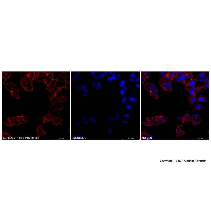
图1. HepG2细胞固定通透后使用 一步法 LumiDye™ 555 鬼笔环肽/核蓝双染试剂盒后共聚焦微镜拍照
常温孵育染色后,LumiDye™555 Phalloidin染色的HepG2可特异性结合细胞微丝(F-肌动蛋白),微丝纤维结构完整、排布清晰;Nucleiblue靶向标记细胞核,核形态规整。融合图像信号分区明确,无明显非特异染色与荧光串色,双染显色均衡。
注:实际检测结果可能因检测仪器、操作条件、细胞类型等差异产生不同,本示例仅提供参考。
常见疑问与解答:
1. 问:鬼笔环肽染色细胞骨架过程中,能使用甲醇固定细胞吗?
答:甲醇会破坏Actin蛋白,因此不能使用含有甲醇的固定液,并且使用不含甲醇的甲醛。
2. 问:本产品是否可以用于染色石蜡切片?
答:不可以。有石蜡切片中的包埋试剂会对肌动蛋白F-actin造成破坏,会出现染色不上的情况。
3. 问:活细胞样本经固定后未进行通透可以染上骨架,但只能染上部分细胞是为什么?
答:经4%多聚甲醛固定的细胞,虽细胞形态和结构仍可保持,但因固定剂的溶脂特性导致细胞膜的通透性发生改变,所以导致LumiDye™标记鬼笔环肽可以进入到细胞对F-actin进行染色。又因未进行TritonX-100通透,所以导致部分细胞被染上。解决方法是:固定且通透。
One-Step LumiDye™ 555 Phalloidin/Nucleiblue Double Staining Kit is a ready-to-use kit that enables specific dual labeling of F-actin and cell nuclei simultaneously. Its core components are LumiDye™ Phalloidin and Nucleiblue, engineered to meet experimental requirements for co-localization analysis of the cytoskeleton and cell nuclei. Core Working Principles:
1. As a phalloidin derivative, LumiDye™ Phalloidin binds specifically and with high affinity to filamentous actin (F-actin) in eukaryotic cells, with no binding to monomeric G-actin. It precisely labels the microfilament structures of the cytoskeleton.
2. Nucleiblue is a highly specific nuclear fluorescent dye that selectively binds to nuclear DNA for distinct fluorescent labeling of cell nuclei. Combined application of the two reagents achieves synchronous staining of F-actin and nuclei.
Unlike conventional separately prepared actin and nuclear staining reagents, this kit adopts a ready-to-use pre-mixed system. Researchers are spared the steps of self-dilution and formulation, drastically cutting experimental operation time. The optimized reagent ratio minimizes non-specific staining to negligible levels. In addition, LumiDye™ Phalloidin features a low molecular weight; its binding to F-actin does not interfere with interactions between actin-binding proteins, thereby preserving the biological functions of F-actin. LumiDye™ dyes represent a new generation series independently developed by our company. Compared with alternative fluorescent dyes, they deliver comprehensive superiorities in brightness, photostability and water solubility. Specifically, LumiDye™ Phalloidin stabilizes F-actin structures and prevents depolymerization to fully preserve intact microfilament morphology. It exhibits no notable disparity in binding affinity toward F-actin across different species and barely disrupts F-actin functionality. Labeled actin filaments retain normal physiological functions: stained muscle fibers remain contractile, and tagged myosin maintains motility. It binds actin subunits at a 1:1 stoichiometric ratio with comparable affinity for thick and thin microfilaments in diverse plant and animal cells, supporting direct quantitative analysis of cellular F-actin. Nucleiblue produces intensely bright fluorescent signals with robust photostability and ultra-low background interference, enabling clear visualization of nuclear morphology, location and numerical variation. These multifold advantages facilitate efficient qualitative and quantitative analysis. This kit is widely compatible with observation under fluorescence microscopes (including laser scanning confocal microscopes) and flow cytometry detection. It suits experimental scenarios such as fundamental cell biology research and cytoskeleton dynamics analysis.
As a pivotal component of the cytoskeleton, filamentous F-actin’s morphology and distribution are tightly correlated with structural alterations of cell nuclei, serving as critical biomarkers for cell migration and morphological remodeling. Phalloidin has long been a gold-standard tool for actin staining due to its specific F-actin binding capacity, while Nucleiblue is a novel high-specificity nuclear fluorescent stain and a top-choice reagent for nuclear labeling. Co-staining with the two constitutes a core analytical method for cellular morphology and subcellular structure assessment.
Against this scientific backdrop, our kit integrates optimized formulations of LumiDye™ Phalloidin and Nucleiblue. LumiDye™ Phalloidin combines precise labeling, intact functional retention and compatibility with quantification. Under fluorescence microscopy, it clearly visualizes the spatial relationship between F-actin microfilament distribution and nuclei for accurate subcellular co-localization analysis. In flow cytometry, it allows simultaneous precise quantification of F-actin expression levels. For drug screening studies, it rapidly detects cytoskeleton remodeling and nuclear morphological shifts post-drug treatment, providing intuitive morphological evidence for investigating drug mechanisms of action. In basic cell biology research, it characterizes dynamic changes in F-actin and nuclei during cell migration, invasion and differentiation, allowing investigations while preserving F-actin physiological functions to maximally restore the native cellular physiological state. Boasting core strengths of high specificity, exceptional stability, superior user-friendliness and full functional preservation, this product provides researchers with an efficient, reliable dual-labeling tool for cytoskeleton-nucleus analysis.
Application Scope:
Labeling of F-actin in cells, frozen tissue sections, yeast and fungi; compatible with immunofluorescence staining.
Product Features:
⁕ Integrated dual labeling, ready-to-use formulation
Premixed staining reagents of LumiDye™ Phalloidin and Nucleiblue for direct use upon opening. No dilution, blending or titration required. One-step staining for both F-actin and cell nuclei, greatly simplifying experimental procedures.
⁕ Precise signals, accurate co-localization
LumiDye™ Phalloidin binds F-actin with high specificity and does not recognize G-actin; Nucleiblue labels cell nuclei accurately with bright signals and low background.
⁕ Broad applicability, beginner-friendly
Time-saving and labor-efficient with reduced experimental steps and reagent consumption. Easy operation for new users. Compatible with coverslip cultures, routine microscopic observation, batch samples, 96-well plates, high-content screening and automated experimental platforms, suitable for high-throughput assays and all routine research experiments.
⁕ Species-independent binding
Unlike antibodies, phalloidin exhibits no significant difference in binding affinity to F-actin across various species, with negligible non-specific staining.
⁕ Function preservation, quantitation compatible
Hardly interferes with the physiological functions of F-actin. Labeled muscle fibers remain contractile and myosin maintains normal motility. It binds actin subunits at a 1:1 stoichiometric ratio with equal affinity for microfilaments in all cell types, supporting quantitative analysis of F-actin.
⁕ Stable results, accurate imaging
Fixed dye ratio and uniform reaction system minimize batch-to-batch and operator-induced variations, delivering excellent staining reproducibility. Simultaneous staining avoids cell displacement and elution issues caused by sequential incubation, yielding more reliable co-localization data, superior signal-to-noise ratio and uniform, steady imaging performance.
Product Specifications:
Item No.
| Product Name
| 50T | 300T | LumiDye™ Ex/Em | Nucleiblue Ex/Em | | O1524682 | One-Step LumiDye™ 488 Phalloidin/Nucleiblue Double Staining Kit
| 5 mL | 30 mL | 490/515 nm | 345-355/460-470 nm | | O1524683 | One-Step LumiDye™ 555 Phalloidin/Nucleiblue Double Staining Kit
| 5 mL | 30 mL | 555/565 nm | 345-355/460-470 nm | | O1524684 | One-Step LumiDye™ 594 Phalloidin/Nucleiblue Double Staining Kit
| 5 mL | 30 mL | 590/617 nm | 345-355/460-470 nm | | O1524685 | One-Step LumiDye™ 633 Phalloidin/Nucleiblue Double Staining Kit
| 5 mL | 30 mL | 630/650 nm | 345-355/460-470 nm | | O1524686 | One-Step LumiDye™ 680 Phalloidin/Nucleiblue Double Staining Kit
| 5 mL | 30 mL | 681/698 nm | 345-355/460-470 nm | | O1524687 | One-Step Rhodamine Phalloidin/Nucleiblue Double Staining Kit
| 5 mL | 30 mL | 546/575 nm | 345-355/460-470 nm |
Note: The usable times of the kit refer to the total number of microwells that can be tested for adherent cells via fluorescence microscopy in 96-well plates.
|
Precautions:
⁕ Phalloidin is toxic (LD₅₀ = 2 mg/kg body weight). Adequate personal protection must be worn during handling.
⁕ Single-staining control groups are recommended for experiments to adjust compensation in flow cytometry assays.
⁕ Fluorescent dyes contained in the kit require storage away from light.
⁕ Phalloidin staining is recommended to be performed after immunolabeling.
⁕ For liquid-cultured yeast, cells in logarithmic growth phase yield far better staining results than those in stationary phase.
⁕ This product is for research use only and shall not be stored in residential premises.
⁕ Follow standard laboratory safety protocols in your facility for your personal safety and health.
Instructions for Use:
I. Pre-experiment Preparation
1. Reagent Preparation
Allow all kit components to equilibrate to room temperature; prepare sufficient PBS buffer, 4% paraformaldehyde and Triton X-100.
2. Instrument Preparation
Fluorescence microscope: Excitation/Emission wavelengths (Ex/Em): 490/515 nm, 555/565 nm, 590/617 nm, 630/650 nm, 681/698 nm, 546/575 nm, 345–355/460–470 nm.
Flow cytometer: Detection channel Excitation/Emission wavelengths (Ex/Em): 490/515 nm, 555/565 nm, 590/617 nm, 630/650 nm, 681/698 nm, 546/575 nm, 345–355/460–470 nm.
3. Cell Preparation
Treat cells with corresponding drugs in accordance with your experimental design.
II. Operating Procedures
Protocol 1: Fluorescence Microscopy Detection (for Suspension Cells)
1. Cell Collection and Washing
(1) Transfer the suspension cell sample to be tested into a centrifuge tube.
(2) Centrifuge at 1000 rpm for 5 minutes at room temperature, then carefully aspirate and discard the supernatant.
2. Cell Resuspension and Counting
(1) Resuspend cells with PBS and perform cell counting.
(2) Transfer 5×10⁵ cells into a new centrifuge tube, centrifuge at low speed of 1000 rpm for 5 minutes, and remove PBS.
Note: Cell density is not strictly restricted for microscopy detection; only ensure consistent cell density between experimental and control groups. Adjust flexibly according to sample type and experimental conditions to achieve clear, distinguishable cell distribution under the microscope field of view.
3. Cell Fixation
(1) Add an appropriate volume of 4% paraformaldehyde fixative, fix cells at room temperature for 15 min, then wash with PBS 2–3 times.
Note: Methanol and other organic solvents will disrupt actin structures during fixation. Methanol-free paraformaldehyde is recommended as the fixative.
4. Cell Permeabilization
(1) (Optional) Permeabilize cells with 0.4% Triton X-100 solution prepared in PBS at room temperature for 15 min to enhance membrane permeability.
(2) Wash with PBS 2–3 times.
Note: Higher centrifugation speed can be adopted when collecting fixed and permeabilized cells.
5. Cell Staining
(1) Add 500 μL of staining working solution (One-Step LumiDye™ 555 Phalloidin/Nucleiblue Double Staining Kit).
Sample Quantity
| 1 | 5 | 10 | One-Step LumiDye™ 555 Phalloidin/Nucleiblue Double Staining Kit
| 500 μL
| 2500 μL
| 5000 μL |
|
(2) Incubate for 15 minutes at room temperature protected from light (incubation time may be optimized based on preliminary trial results).
Note: Incubation duration and loading volume of the dye can be adjusted according to different cell types, preliminary experimental data, or customized dosages set by experimental conditions.
6. Washing
(1) Aspirate and discard the staining working solution after incubation completes.
(2) Wash the cells with PBS 2–3 times.
7. Microscopic Observation and Imaging
(1) 96-well plate method: Add 100 μL cell suspension into a 96-well plate, let stand for a short while until cells settle naturally to the bottom, then observe under a fluorescence microscope.
Note: The volume of cell suspension can be adjusted according to cell counts; optimal cell confluency in the microscope field is 70%–80%.
(2) Glass slide method: Pipette 20–30 μL cell suspension onto a clean glass slide, gently place a coverslip to avoid air bubbles, then observe under a fluorescence microscope.
(3) Locate target fields and capture images separately under preset filter sets.
Protocol 2: Fluorescence Microscopy Detection (for Adherent Cells)
1. Cell Treatment
(1) Seed cells into well plates one day in advance to reach 70%–85% confluence. After cells adhere fully, treat them following your experimental design.
2. Cell Washing
(1) Aspirate cell culture medium directly from the culture plate.
(2) Gently wash cells 2–3 times with PBS at volume matched to plate format (e.g., 100 μL per well for 96-well plates, 150 μL per well for 48-well plates, 250 μL per well for 24-well plates, 500 μL per well for 12-well plates, 1 mL per well for 6-well plates). Aspirate all PBS after washing.
3. Cell Fixation
(1) Add an appropriate volume of 4% paraformaldehyde fixative, fix cells at room temperature for 15 min, then rinse with PBS 2–3 times.
Note: Methanol and other organic solvents disrupt actin structures during fixation. Methanol-free paraformaldehyde is recommended as fixative.
4. Cell Permeabilization
(1) (Optional) Permeabilize cells with 0.4% Triton X-100 in PBS at room temperature for 15 min to boost membrane permeability.
(2) Rinse with PBS 2–3 times.
Cell Staining
(1) Add staining working solution (One-Step LumiDye™ 555 Phalloidin/Nucleiblue Double Staining Kit) to each well at the same volume specified in Step 2 (e.g., 100 μL/well for 96-well, 150 μL/well for 48-well, 250 μL/well for 24-well, 500 μL/well for 12-well, 1 mL/well for 6-well plates).
Sample Quantity (96-well Plate)
| 1 | 5 | 10 | One-Step LumiDye™ 555 Phalloidin/Nucleiblue Double Staining Kit
| 100 μL
| 500 μL
| 1000 μL |
|
(2) Incubate for 15 minutes at room temperature in the dark (incubation time can be optimized based on preliminary experimental results).
Note: The incubation duration and loading volume of the dye can be adjusted according to different cell types, preliminary test data, or customized dosages for experimental conditions.
6. Washing
(1) Aspirate the staining working solution from each well once incubation is finished.
(2) Wash the cells 2–3 times with PBS at the same volume indicated in Step 2.
7. Microscopic Observation and Image Capture
(1) The labeled phalloidin exhibits excellent photostability. Samples can be imaged directly in PBS; for optimal performance, an anti-fade mounting medium is also applicable.
Note: Imaging immediately is strongly recommended if samples are not mounted. For best outcomes, stained samples should be mounted with suitable mounting medium and stored at 4 °C protected from light.
Protocol 3: Flow Cytometry Detection (for Suspension and Adherent Cells)
1. Collection and Preparation of Single-Cell Suspension
⁕ Suspension cells: Follow the procedures in Protocol 1; finally collect 4×10⁵–6×10⁵ cells.
⁕ Adherent cells:
a. Digest cells with an appropriate amount of trypsin digestion solution.
Note: The dosage of trypsin, digestion duration and temperature shall be operated per laboratory routine experience.
b. Key step: Add complete cell culture medium to terminate digestion.
c. Transfer cell suspension into a suitable centrifuge tube, centrifuge at 1000 rpm for 5 min at room temperature, then aspirate and discard supernatant.
d. Resuspend cells with PBS and conduct cell counting.
e. Take 4×10⁵–6×10⁵ cells, centrifuge at 1000 rpm for 5 min at room temperature, remove supernatant.
f. Wash cells with PBS, centrifuge at 1000 rpm for 5 min at room temperature, discard supernatant.
2. Cell Fixation
(1) Add appropriate volume of 4% paraformaldehyde fixative, fix cells at room temperature for 15 min, then wash with PBS 2–3 times.
Note: Methanol and other organic solvents will disrupt actin structures during fixation. Methanol-free paraformaldehyde is recommended as fixative.
3. Cell Permeabilization
(1) (Optional) Permeabilize cells with 0.4% Triton X-100 diluted in PBS at room temperature for 15 min to enhance membrane permeability.
(2) Rinse with PBS 2–3 times.
Note: Higher centrifugation speed can be used when harvesting fixed and permeabilized cells.
4. Cell Staining
(1) Add 0.5 mL staining working solution to resuspend into single-cell suspension. Incubate for 15 min at room temperature away from light.
Note: Washing with PBS 1–3 times is also feasible after incubation.
Sample Quantity
| 1 | 5 | 10 | One-Step LumiDye™ 555 Phalloidin/Nucleiblue Double Staining Kit
| 500 μL
| 2500 μL
| 5000 μL |
Note: The incubation time and loading volume of the dye can be adjusted according to different cell types used in the experiment, preliminary experimental results or customized dosages set for experimental conditions. |
5. Detection
(1) Important: Perform flow cytometry detection within 1 hour after staining to ensure stable fluorescent signals.
(2) Analyze samples via flow cytometer under preset detection channels.
(3) Adjust voltage using blank control (unstained cells) first to place cell populations at the lower-left quadrant of the coordinate system.
(4) Subsequently test negative and positive controls to verify valid experimental system.
(5) Finally test experimental samples, record and analyze the mean fluorescence intensity of cells.
Protocol 4: Fluorescence Microscopy Detection (for Tissue Sections)
1. Section Washing
(1) Rinse tissue sections twice with PBS.
2. Fixation
(1) Fix tissue sections or cells with 3.7% formaldehyde solution prepared in PBS at room temperature for approximately 10–20 min.
Note: Methanol destroys actin structure; fixatives containing methanol are prohibited, and methanol-free formaldehyde is strongly recommended.
(2) Wash 2–4 times with PBS containing 0.1% Triton X-100, 5 minutes per wash.
3. Staining and Washing
(1) Drop staining working solution onto each section at a volume of 200 µL per slide, incubate for 30–60 min at room temperature protected from light. Place slides in a slide staining box during incubation to prevent liquid evaporation.
Sample Quantity
| 1 | 5 | 10 | One-Step LumiDye™ 555 Phalloidin/Nucleiblue Double Staining Kit
| 200 μL
| 1000 μL
| 2000 μL |
Note: The incubation time and volume of the dye applied can be adjusted according to different types of tissue sections, preliminary experimental results, or customized dosages specified for experimental conditions. |
(2) Wash 2–4 times with PBS containing 0.1% Triton X-100, 5 minutes each time.
4. Microscopic Observation and Image Capture
(1) Samples can then be directly observed under a fluorescence microscope.
Note: For long-term preservation, mount the slides after air-drying naturally and store at 4 °C away from light; the preservation period can reach approximately 6 months.
III. Result Interpretation
1. Qualitative Analysis (Microscopy)

Figure 1. Fluorescence micrograph of fixed and permeabilized HeLa cells co-stained with LumiDye™ 555 Phalloidin and Hoechst 33342
After staining incubation at room temperature, LumiDye™ 555 Phalloidin specifically binds to cellular microfilaments (F-actin) in frozen mouse lung sections, presenting intact microfilament fibers with distinct and orderly arrangement. Nucleiblue targets and labels cell nuclei with regular nuclear morphology. The merged image shows clearly divided signal regions, with negligible non-specific staining and fluorescence crosstalk, and balanced signal intensity for dual staining.
Note: Actual test results may vary depending on detection equipment, operating conditions, cell types and other factors. This example is for reference only.
Frequently Asked Questions:
1. Q: Can methanol be used for cell fixation during phalloidin cytoskeleton staining?
A: Methanol degrades actin protein. Fixatives containing methanol must not be used; methanol-free paraformaldehyde is required.
2. Q: Can this product be used for staining paraffin sections?
A: No. Embedding agents in paraffin sections damage F-actin, which will cause poor or absent staining signals.
3. Q: Cytoskeleton staining is partially successful in fixed live cell samples without permeabilization. What causes this partial staining?
A: Cells fixed with 4% paraformaldehyde maintain intact cellular morphology and structure, yet the lipolytic property of the fixative modifies cell membrane permeability, allowing LumiDye™-conjugated phalloidin to penetrate cells and bind F-actin. However, the lack of Triton X-100 permeabilization limits uniform penetration, resulting in only partial cell staining. The solution is to perform both fixation and permeabilization procedures.

 ZJ26F0737962


 危险品化学品经营许可证(带存储)