Receptor tyrosine kinase binding ligands of the EGF family and activating several signaling cascades to convert extracellular cues into appropriate cellular responses. Known ligands include EGF, TGFA/TGF-alpha, amphiregulin, epigen/EPGN, BTC/betacellulin, epiregulin/EREG and HBEGF/heparin-binding EGF. Ligand binding triggers receptor homo- and/or heterodimerization and autophosphorylation on key cytoplasmic residues. The phosphorylated receptor recruits adapter proteins like GRB2 which in turn activates complex downstream signaling cascades. Activates at least 4 major downstream signaling cascades including the RAS-RAF-MEK-ERK, PI3 kinase-AKT, PLCgamma-PKC and STATs modules. May also activate the NF-kappa-B signaling cascade. Also directly phosphorylates other proteins like RGS16, activating its GTPase activity and probably coupling the EGF receptor signaling to the G protein-coupled receptor signaling. Also phosphorylates MUC1 and increases its interaction with SRC and CTNNB1/beta-catenin. Isoform 2 may act as an antagonist of EGF action.
宿主种属
小鼠(Mouse)
特异性
EGFR
种属反应性
人(Human),小鼠(Mouse)
免疫原
Recombinant EGFR expressed in E.coli (AA 888-1210)
阳性对照
WB: A431, HeLa, MCF7, A549, NIH/3T3 and HUVEC cell lysates. IHC: Human kidney, placent, lung cancer, stomach, adrenal gland, liver cancer tissues. IF/ICC: HeLa cells.
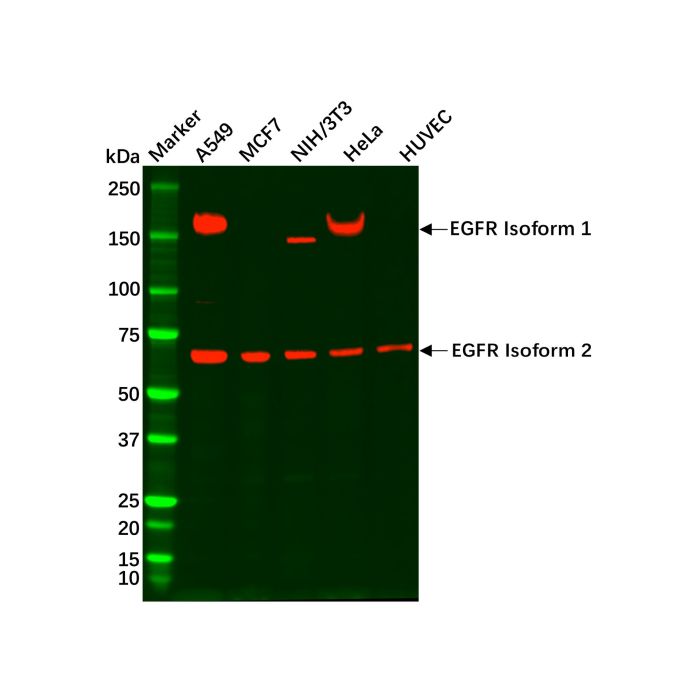
EGFR Mouse mAb (Ab101163) - Western Blot All lanes: EGFR Mouse mAb (Ab101163) at 1/1000 dilution Samples: Lysates at 20 µg per lane Secondary: Goat Anti-Mouse IgG H&L (HRP) (Ab138040) at 1/20000 dilution
Predicted band size: 175 kDa Observed band size: 169 & 69 kDa Exposure time: 10.4 s
EGFR Mouse mAb (Ab101163) - Western Blot All lanes: EGFR Mouse mAb (Ab101163) at 1/1000 dilution Samples: Lysates at 20 µg per lane Secondary: Goat Anti-Mouse IgG H&L (HRP) (Ab138040) at 1/20000 dilution
Predicted band size: 175 kDa Observed band size: 169 & 69 kDa Exposure time: 2.3 s
EGFR Mouse mAb (Ab101163) - IHC DAB staining on IHC Sample: Human Kidney Tissue Primary Ab: 5 μg/mL EGFR Mouse mAb (Ab101163) Second Ab: 2 µg/mL HRP-Linked Caprine Anti-Mouse IgG Polyclonal Antibody.
EGFR Mouse mAb (Ab101163) - IHC DAB staining on IHC Sample: Human Placenta Tissue Primary Ab: 5 μg/mL EGFR Mouse mAb (Ab101163) Second Ab: 2 µg/mL HRP-Linked Caprine Anti-Mouse IgG Polyclonal Antibody.
EGFR Mouse mAb (Ab101163) - IHC DAB staining on IHC Sample: Human Lung cancer Tissue Primary Ab: 5 μg/mL EGFR Mouse mAb (Ab101163) Second Ab: 2 µg/mL HRP-Linked Caprine Anti-Mouse IgG Polyclonal Antibody.
EGFR Mouse mAb (Ab101163) - IHC DAB staining on IHC Sample: Human Stomach Tissue Primary Ab: 5 μg/mL EGFR Mouse mAb (Ab101163) Second Ab: 2 µg/mL HRP-Linked Caprine Anti-Mouse IgG Polyclonal Antibody.
EGFR Mouse mAb (Ab101163) - IHC DAB staining on IHC Sample: Human Adrenal gland Tissue Primary Ab: 5 μg/mL EGFR Mouse mAb (Ab101163) Second Ab: 2 µg/mL HRP-Linked Caprine Anti-Mouse IgG Polyclonal Antibody.
EGFR Mouse mAb (Ab101163) - IHC DAB staining on IHC Sample: Human Liver cancer Tissue Primary Ab: 5 μg/mL EGFR Mouse mAb (Ab101163) Second Ab: 2 µg/mL HRP-Linked Caprine Anti-Mouse IgG Polyclonal Antibody.
EGFR Mouse mAb (Ab101163) - IF/ICC IF analysis of EGFR Mouse mAb (Ab101163) in HeLa cells. The cells were fixed and permeabilized with 100% methanol for 5 minutes, and blocked with 5% BSA for 1 hour at room temperature. Cells were stained with EGFR Mouse mAb (Ab101163) at 1/200 dilution in blocking buffer overnight at 4°C, and then incubated with Goat Anti-Mouse IgG H&L (FITC) (Ab179003) at a dilution of 1/1000 for 1 hour at room temperature in the dark (green). Cells were counterstained with DAPI (blue).



 ZJ24F0404291


 危险品化学品经营许可证(带存储)