线粒体是真核细胞的 "能量工厂",通过氧化磷酸化产生 ATP 为细胞生命活动提供能量。线粒体膜电位 (ΔΨm) 是指线粒体内膜两侧形成的外正内负的电位差,正常生理状态下约为 - 180 mV。它的形成与维持对线粒体的能量代谢、物质运输、钙离子稳态调节及细胞生存至关重要,是反映线粒体功能状态最敏感的指标之一。
线粒体膜电位下降是细胞凋亡早期的标志性事件,远早于细胞形态学变化、Caspase 激活和 DNA 断裂。当细胞受到凋亡诱导剂、氧化应激、毒素等刺激时,线粒体通透性转换孔 (MPTP) 开放,导致质子梯度消散,膜电位去极化,最终触发细胞凋亡程序。因此,检测线粒体膜电位变化成为研究细胞凋亡机制、评估药物毒性和筛选线粒体靶向药物的重要手段。
JC-1 是一种亲脂性阳离子荧光染料,其荧光特性与线粒体膜电位(MMP)直接相关,具有电位依赖性跨膜聚集特性,荧光信号随线粒体膜电位变化发生特异性改变:
健康细胞线粒体膜电位高:JC-1 聚集在线粒体基质中形成J - 1聚集体(aggregate),JC-1多聚体的最大激发波长为585 nm,最大发射波长为590 nm,呈现红色荧光;
凋亡 / 损伤细胞线粒体膜电位下降:JC-1 无法聚集,以单体(monomer)形式分散在胞质中,JC-1单体的最大激发波长为514 nm,最大发射波长为529 nm,呈现绿色荧光;
通过红 / 绿荧光强度比值,可精准定量线粒体膜电位变化,排除细胞数量、染色效率等无关变量干扰。实际观察时,常规设置红色荧光和绿色荧光即可。
M1508380 | 组分 | 100 T | 储存条件 | M1508380A | JC-1(500×) | 4×50 μL | -20℃. 避光保存. | M1508380B | CCCP(10 mM) | 100 μL | -20℃. 避光保存. | M1508380C | 缓冲液(5×) | 100 mL | 2-8℃. |
|
使用方法
1. 试剂配制
JC-1工作液配制:六孔板每孔所需JC-1染色工作液的量为1 mL,其它培养器皿的JC-1染色工作液的用量以此类推;对于细胞悬液每50-100万细胞需0.5 mL JC-1染色工作液。取适量JC-1(500×),按照每20 µL JC-1(500×)加入8 mL超纯水的比例稀释JC-1。充分溶解并混匀JC-1。然后再加入2 mL JC-1染色缓冲液(5×),混匀后即为JC-1染色工作液。
必须先把JC-1(500×)用超纯水充分溶解混匀后,才可以加入JC-1染色缓冲液(5×)。不可先配制JC-1染色缓冲液(1×)再加入JC-1(500×),这样JC-1会很难充分溶解,会严重影响后续的检测。
2. 阳性对照的设置
把试剂盒中提供的CCCP(10 mM)推荐按照1:200的比例加入到细胞培养液中,稀释至50 µM,处理细胞20分钟。随后按照下述方法装载JC-1,进行线粒体膜电位的检测。对于大多数细胞,通常50 µM CCCP处理20分钟后线粒体的膜电位会完全丧失,JC-1染色后观察应呈绿色荧光;而正常的细胞经JC-1染色后应显示红色荧光。对于不同的细胞,CCCP的作用浓度和作用时间可能有所不同,需自行参考相关文献资料确定。
3. 对于悬浮细胞:
(1)取10-60万细胞,重悬于0.5 mL细胞培养液中,细胞培养液中可以含血清和酚红。
(2)加入0.5 mL JC-1染色工作液,颠倒数次混匀。细胞培养箱中37ºC孵育20分钟。
(3)孵育结束后,600 g ,4ºC离心5分钟,沉淀细胞,弃上清。注意尽量不要吸除细胞。
(4)用JC-1染色缓冲液(1×)洗涤2次:加入1 mL JC-1染色缓冲液(1×)重悬细胞,600 g, 4ºC离心5分钟,沉淀细胞,弃上清。再加入1 mL JC-1染色缓冲液(1×)重悬细胞,600 g ,4ºC离心5分钟,沉淀细胞,弃上清。
(5)再用适量JC-1染色缓冲液(1×)重悬后,用荧光显微镜或激光共聚焦显微镜观察,也可以用荧光分光光度计检测或流式细胞仪分析。
4. 对于贴壁细胞:
注意:对于贴壁细胞,如果希望采用荧光分光光度计或流式细胞仪检测,可以先收集细胞,重悬后参考悬浮细胞的检测方法。
(1) 对于六孔板的一个孔,吸除培养液,根据具体实验如有必要可以用PBS或其它适当溶液洗涤细胞一次,加入1mL细胞培养液。细胞培养液中可以含有血清和酚红。
(2) 加入1mL JC-1染色工作液,充分混匀。细胞培养箱中37ºC孵育20分钟。
(3) 在孵育期间,按照每1mL JC-1染色缓冲液(5×)加入4mL蒸馏水的比例,配制适量的JC-1染色缓冲液(1×),并放置于冰浴。
(4) 37ºC孵育结束后, 吸除上清,用JC-1染色缓冲液(1×)洗涤2次。
(5) 加入2mL JC-1染色缓冲液(1×)后,可用荧光显微镜或激光共聚焦显微镜下观察。
5. 对于纯化的线粒体:
(1) 把配制好的JC-1染色工作液再用JC-1染色缓冲液(1×)稀释5倍。
(2) 0.9 mL 5倍稀释的JC-1染色工作液中加入0.1 mL总蛋白量为10-100 µg纯化的线粒体。
(3) 用荧光分光光度计或荧光酶标仪检测:混匀后直接用荧光分光光度计进行时间扫描(time scan),激发波长为485 nm,发射波长为590 nm。如果使用荧光酶标仪,激发波长不能设置为485 nm时,可以在475-520 nm范围内设置激发波长。另外,也可以参考下面步骤6中的波长设置进行荧光检测。
(4) 用荧光显微镜或激光共聚焦显微镜观察:方法同下面的步骤6。
6. 荧光观测和结果分析
检测JC-1单体时可以把激发光设置为490 nm,发射光设置为530 nm;检测JC-1聚合物时,可以把激发光设置为525 nm,发射光设置为590 nm。注意:此处测定荧光时不必把激发光和发射光设置在最大激发波长和最大发射波长。如使用荧光显微镜观察,检测JC-1单体时可以参考观察其它绿色荧光时的设置,如观察GFP或FITC时的设置;检测JC-1聚合物时可以参考观察其它红色荧光,如碘化丙啶或Cy3时的设置。
注意事项
(1) JC-1(500×)在4ºC、冰浴等较低温度情况下会凝固而粘在离心管管底、管壁或管盖内,可以20-25ºC水浴温育片刻至全部融解后使用。
(2) 装载完JC-1后用JC-1染色缓冲液(1×)洗涤时,使JC-1染色缓冲液(1×)保持4ºC左右,此时的洗涤效果较好。
(3) 为防止荧光淬灭,操作和存放过程中需尽量注意避光,JC-1探针装载完并洗涤后尽量在30分钟内完成后续检测。在检测前需冰浴保存。
(4) 如果发现JC-1染色缓冲液(5×)中有沉淀,必须全部溶解后才能使用,为促进溶解可以在37ºC加热。
(5) CCCP为线粒体电子传递链抑制剂,对人体有害,操作时请小心,并注意有效防护以避免直接接触人体或吸入体内。
(6) 本产品仅限于专业人员的科学研究用,不得用于临床诊断或治疗,不得用于食品或药品,为了您的安全和健康,请穿实验服并戴一次性手套操作。

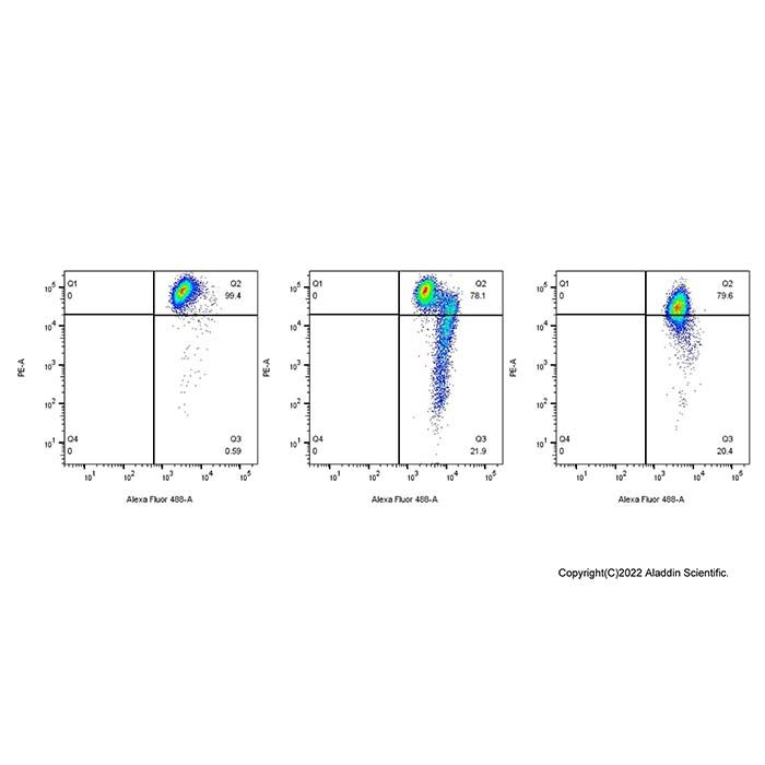
图1:本试剂盒检测Jurkat细胞凋亡效果如图所示:正常细胞(左)存在少量凋亡,表现为出现少量线粒体膜电位崩塌细胞;诱导凋亡细胞(中,10 μM喜树碱处理Jurkat细胞4 h)出现大量线粒体膜电位崩塌细胞;CCCP处理细胞(右,阳性对照)几乎所有细胞线粒体膜电位崩塌。

图2:本试剂盒检测HT-29细胞线粒体膜电位的效果图。正常的HT-29细胞线粒体中JC-1以聚合物形式存在,呈明亮的红色荧光,细胞中的绿色荧光非常弱。

图3:本试剂盒检测CCCP处理后HT-29细胞线粒体膜电位的效果图。CCCP处理的的HT-29细胞线粒体中JC-1以单体形式存在,红色荧光显著减少,绿色荧光显著增加。
Mitochondria are the "powerhouse" of eukaryotic cells, producing ATP through oxidative phosphorylation to provide energy for cellular life activities. Mitochondrial membrane potential (ΔΨm) refers to the potential difference with positive outside and negative inside formed across the inner mitochondrial membrane, which is approximately -180 mV under normal physiological conditions. Its formation and maintenance are crucial for mitochondrial energy metabolism, substance transport, calcium homeostasis regulation and cell survival, and it is one of the most sensitive indicators reflecting mitochondrial functional status.
A decrease in mitochondrial membrane potential is a hallmark event in the early stage of apoptosis, occurring much earlier than morphological changes, Caspase activation and DNA fragmentation. When cells are stimulated by apoptosis inducers, oxidative stress, toxins, etc., the mitochondrial permeability transition pore (MPTP) opens, leading to the dissipation of the proton gradient, membrane potential depolarization, and ultimately triggering the apoptotic program. Therefore, detecting changes in mitochondrial membrane potential has become an important means for studying the mechanism of apoptosis, evaluating drug toxicity and screening mitochondria-targeted drugs.
JC-1 is a lipophilic cationic fluorescent dye whose fluorescence properties are directly related to mitochondrial membrane potential (MMP). It has potential-dependent transmembrane aggregation characteristics, and its fluorescence signal changes specifically with mitochondrial membrane potential:
High mitochondrial membrane potential in healthy cells: JC-1 accumulates in the mitochondrial matrix to form J-aggregates. The maximum excitation wavelength of JC-1 aggregates is 585 nm, and the maximum emission wavelength is 590 nm, showing red fluorescence.
Decreased mitochondrial membrane potential in apoptotic/damaged cells: JC-1 cannot aggregate and exists as monomers dispersed in the cytoplasm. The maximum excitation wavelength of JC-1 monomers is 514 nm, and the maximum emission wavelength is 529 nm, showing green fluorescence.
The red/green fluorescence intensity ratio can accurately quantify changes in mitochondrial membrane potential and eliminate interference from irrelevant variables such as cell number and staining efficiency. For actual observation, conventional settings for red fluorescence and green fluorescence are sufficient.
Kit Components
M1508380 | Component | 100 T | Storage conditions | M1508380A | JC-1(500×) | 4×50 μL | -20℃. Store in the dark | M1508380B | CCCP(10 mM) | 100 μL | -20℃. Store in the dark | M1508380C | Assay Buffer (5×) | 100 mL | 2-8℃. |
|
Protocol
1.Reagent Preparation
Preparation of JC-1 Working Solution: The volume of JC-1 staining working solution required per well of a 6-well plate is 1 mL, and the volume for other culture vessels can be calculated proportionally. For cell suspensions, 0.5 mL of JC-1 staining working solution is needed for every 500,000 to 1,000,000 cells. Take an appropriate amount of JC-1 (500×), dilute it by adding 8 mL of ultrapure water per 20 μL of JC-1 (500×). Fully dissolve and mix JC-1. Then add 2 mL of JC-1 Staining Buffer (5×) and mix well to obtain the JC-1 staining working solution.
Important Note: JC-1 (500×) must be fully dissolved and mixed with ultrapure water first before adding JC-1 Staining Buffer (5×). Do not prepare JC-1 Staining Buffer (1×) first and then add JC-1 (500×), as this will make it difficult for JC-1 to dissolve completely and seriously affect subsequent detection.
2. Setting Up the Positive Control
Dilute the CCCP (10 mM) provided in the kit at a recommended ratio of 1:200 into the cell culture medium to a final concentration of 50 μM, and treat the cells for 20 minutes. Then load JC-1 according to the following methods to detect mitochondrial membrane potential. For most cells, treatment with 50 μM CCCP for 20 minutes will completely abolish mitochondrial membrane potential, and cells should show green fluorescence after JC-1 staining; while normal cells should show red fluorescence after JC-1 staining. For different cell types, the optimal concentration and incubation time of CCCP may vary, please refer to relevant literature for determination.
3. For Suspension Cells
(1) Take 100,000 to 600,000 cells and resuspend them in 0.5 mL of cell culture medium, which may contain serum and phenol red.
(2) Add 0.5 mL of JC-1 staining working solution and mix by inverting the tube several times. Incubate at 37°C in a cell culture incubator for 20 minutes.
(3) After incubation, centrifuge at 600 g for 5 minutes at 4°C to pellet the cells, and discard the supernatant. Be careful not to aspirate the cells.
(4) Wash twice with JC-1 Staining Buffer (1×): Add 1 mL of JC-1 Staining Buffer (1×) to resuspend the cells, centrifuge at 600 g for 5 minutes at 4°C, pellet the cells and discard the supernatant. Repeat this step once.
(5) Resuspend the cells in an appropriate volume of JC-1 Staining Buffer (1×), then observe with a fluorescence microscope or laser confocal microscope, or detect with a fluorescence spectrophotometer or analyze by flow cytometry.
4. For Adherent Cells
Note: For adherent cells, if detection by fluorescence spectrophotometer or flow cytometry is desired, collect the cells first, resuspend them, and then refer to the detection method for suspension cells.
(1) For one well of a 6-well plate, aspirate the culture medium. If necessary according to the specific experiment, wash the cells once with PBS or other appropriate solution, and add 1 mL of cell culture medium, which may contain serum and phenol red.
(2) Add 1 mL of JC-1 staining working solution and mix thoroughly. Incubate at 37°C in a cell culture incubator for 20 minutes.
(3) During the incubation period, prepare an appropriate amount of JC-1 Staining Buffer (1×) by adding 4 mL of distilled water per 1 mL of JC-1 Staining Buffer (5×), and place it on ice.
(4) After the 37°C incubation, aspirate the supernatant and wash the cells twice with JC-1 Staining Buffer (1×).
(5) Add 2 mL of JC-1 Staining Buffer (1×), then observe under a fluorescence microscope or laser confocal microscope.
5. For Purified Mitochondria
(1) Dilute the prepared JC-1 staining working solution 5-fold with JC-1 Staining Buffer (1×).
(2) Add 0.1 mL of purified mitochondria with a total protein amount of 10-100 μg to 0.9 mL of the 5-fold diluted JC-1 staining working solution.
(3) Detection by fluorescence spectrophotometer or fluorescence microplate reader: After mixing, perform time scan directly with a fluorescence spectrophotometer at an excitation wavelength of 485 nm and an emission wavelength of 590 nm. If using a fluorescence microplate reader and the excitation wavelength cannot be set to 485 nm, the excitation wavelength can be set in the range of 475-520 nm. Alternatively, refer to the wavelength settings in Step 6 below for fluorescence detection.
(4) Observation by fluorescence microscope or laser confocal microscope: Follow the method in Step 6 below.
6. Fluorescence Detection Wavelength Settings
For detecting JC-1 monomers, set the excitation wavelength to 490 nm and the emission wavelength to 530 nm; for detecting JC-1 aggregates, set the excitation wavelength to 525 nm and the emission wavelength to 590 nm.
Note: It is not necessary to set the excitation and emission wavelengths to their maximum values for fluorescence measurement. When using a fluorescence microscope for observation, the settings for detecting JC-1 monomers can refer to those for observing other green fluorescent proteins such as GFP or FITC; the settings for detecting JC-1 aggregates can refer to those for observing other red fluorescent dyes such as propidium iodide or Cy3.
Precautions
(1) JC-1 (500×) may solidify and stick to the bottom, wall or cap of the centrifuge tube at low temperatures such as 4°C or ice bath. Incubate in a 20-25°C water bath for a few minutes until completely thawed before use.
(2) When washing with JC-1 Staining Buffer (1×) after loading JC-1, keep the buffer at approximately 4°C for better washing effect.
(3) To prevent fluorescence quenching, avoid light as much as possible during operation and storage. Complete subsequent detection within 30 minutes after loading JC-1 and washing. Keep samples on ice before detection.
(4) If precipitation is found in JC-1 Staining Buffer (5×), it must be completely dissolved before use. Heating at 37°C can promote dissolution.
(5) CCCP is an inhibitor of the mitochondrial electron transport chain and is harmful to humans. Please handle with caution and take effective protective measures to avoid direct contact with the human body or inhalation.
(6) This product is for scientific research use only by professional personnel. It shall not be used for clinical diagnosis or treatment, nor for food or drugs. For your safety and health, please wear a lab coat and disposable gloves during operation.
Results Display
Figure 1: Apoptosis effect of Jurkat cells detected by this kit. Normal cells (left) show a small amount of apoptosis, manifested as the presence of a few cells with collapsed mitochondrial membrane potential; apoptosis-induced cells (middle, Jurkat cells treated with 10 μM camptothecin for 4 h) show a large number of cells with collapsed mitochondrial membrane potential; CCCP-treated cells (right, positive control) show almost complete collapse of mitochondrial membrane potential in all cells.

Figure 2: Effect of this kit on detecting mitochondrial membrane potential in HT-29 cells. In normal HT-29 cells, JC-1 exists as aggregates in mitochondria, showing bright red fluorescence, and the green fluorescence in cells is very weak.

Figure 3: Effect of this kit on detecting mitochondrial membrane potential in CCCP-treated HT-29 cells. In CCCP-treated HT-29 cells, JC-1 exists as monomers in mitochondria, with significantly reduced red fluorescence and significantly increased green fluorescence.

 H2611253


 危险品化学品经营许可证(带存储)