Introduction: Streptavidin is a tetrameric protein purified from Streptomyces avidinii that binds very tightly to the vitamin biotin with a dissociation constant on the order of ≈10⁻¹⁴ mol/L. The high affinity recognition of biotin and biotinylated molecules has made streptavidin one of the most important components in diagnostics and laboratory kits. TRITC conjugated streptavidin can be used to localize or to detect biotinylated biomolecules in cells, tissue sections or other fluorescent immunoassays. Recommended Usage for Application: IF: 1:1000 WB: 1:4000-1:30000 Since applications vary, you should determine the optimum working dilution for the product that is appropriate for your specific need.
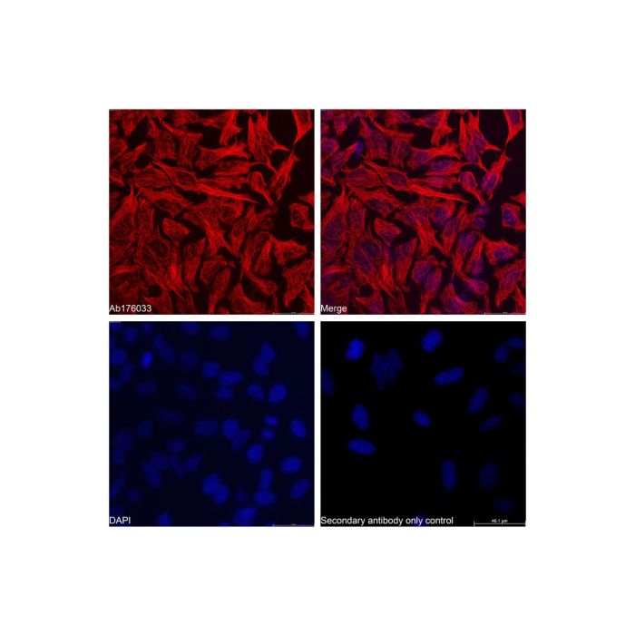
Recombinant Streptavidin Protein (TRITC) (rp169049) - IF IF analysis of beta Tubulin (red) in HeLa cells. The cells were fixed and permeabilized with 100% methanol for 5 minutes, and blocked with 2% BSA for 1 hour at room temperature. Cells were stained with beta Tubulin Mouse mAb (Biotin) (Ab176033) at 1/2000 dilution in blocking buffer overnight at 4°C, and then incubated with Recombinant Streptavidin Protein (TRITC) (rp169049) at a dilution of 1/1000 for 1 hour at room temperature in the dark (red). Cells were counterstained with DAPI (blue). PBS instead of the primary antibody was used as the secondary antibody only control. Images were taken on the confocal laser scanning microscope.
Recombinant Streptavidin Protein (TRITC) (rp169049) - IF IF analysis of beta Actin (red) in HeLa cells. The cells were fixed and permeabilized with 100% methanol for 5 minutes, and blocked with 2% BSA for 1 hour at room temperature. Cells were stained with beta Actin Mouse mAb (Biotin) (Ab175850) at 1/2000 dilution in blocking buffer overnight at 4°C, and then incubated with Recombinant Streptavidin Protein (TRITC) (rp169049) at a dilution of 1/1000 for 1 hour at room temperature in the dark (red). Cells were counterstained with DAPI (blue). PBS instead of the primary antibody was used as the secondary antibody only control. Images were taken on the confocal laser scanning microscope.


 ZJ25F0319103


 危险品化学品经营许可证(带存储)