
计算溶液所需的质量、体积或浓度。
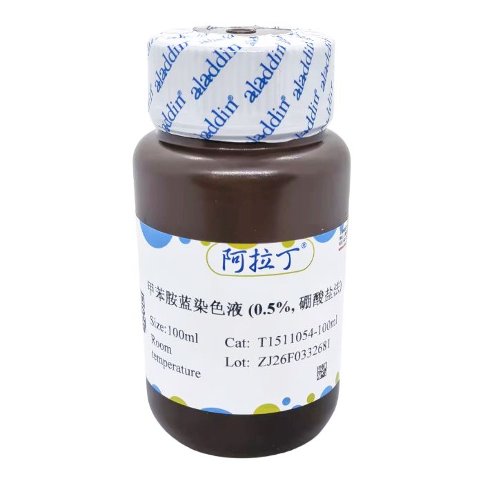
甲苯胺蓝染色液 (0.5%, 硼酸盐法)
有货
BioReagent,生物染色剂,用于显微镜 级 ,适用于对基线干扰要求严格的色谱和分析工作流程。
室温。常规运输 。请查阅批次 COA 获取详细规格。
SDS、COA、产品数据表及规格说明书均可下载。可通过批号查询获取批次 COA。
在色谱分析、有机合成和交叉偶联反应领域已被 0 篇同行评审文献引用。
甲苯胺蓝 (Toluidine Blue O) 是一种常用的人工合成染料,属于醌亚胺染料类。这类染料主要含有胺基和醌型苯环两个发色团,从而成色原显色。甲苯胺蓝中的阳离子有染色作用,组织细胞的酸性物质与其中的阳离子相结合而被染色。
甲苯胺蓝不仅含有两个发色团,还含有两个助色团,能促使染料产生电离成盐类,帮助发色团对组织产生染色力,使切片上的组织细胞着色,可染细胞核使之呈蓝色;肥大细胞胞质内含有肝素和组织胺等异色性物质遇到甲苯胺蓝可呈异染性紫红色。甲苯胺蓝染色液 (Toluidine Blue, 0.5%, 硼酸盐法) 由于硼酸盐缓冲液呈强碱性,更利于组织细胞的着色。
操作步骤(仅供参考):
(一)肥大细胞染色
1、脱蜡至蒸馏水。
2、浸染于甲苯胺蓝染色液10-15min,具体的染色时间根据切片厚度和组织的不同而定。
3、蒸馏水或去离子水轻轻冲洗。
4、(可选)0.5%冰乙酸分化,直到细胞核和颗粒清晰可见。
5、快速95%和无水乙醇脱水。
6、二甲苯透明,封固。
染色结果:肥大细胞呈紫红色;背景呈淡蓝色。
(二)软骨染色
1、石蜡切片入二甲苯2次每次15min。
2、系列乙醇各1min。
3、自来水洗2min。
4、入Toluidine Blue O Stain 浸染30min。
5、自来水洗2min,滤纸吸干水分。
6、丙酮分化至软骨细胞呈紫蓝色清楚可见。
7、逐级乙醇脱水。
8、二甲苯透明,中性树胶封固。
染色结果:软骨、成骨细胞呈紫红色;背景呈淡蓝色。
(三)细胞涂片染色
1、用20%的乙醇溶液稀释甲苯胺蓝染色液,一般要求稀释到0.1%即可。
2、细胞涂片后,立即放入95%的乙醇中固定,取出放在纸巾上。
3、滴加稀释后的甲苯胺蓝染色液进行滴染,加盖玻片让染料渗透到细胞中。
4、将玻片竖起,稍加压力,使多余染料被纸巾吸去。
5、无需干燥,直接镜检。
染色结果:细胞核、淋巴细胞呈深蓝色;核仁呈紫红色;红细胞呈橘红;细胞质、单核细胞呈淡蓝色。
(四)原位杂交染色
1、 用蒸馏水或去离子水稀释到相应的浓度,凭经验一般稀释比例大于1:100。
2、 玻片在稀释好的染色液中短暂浸泡。
3、 在蒸馏水或去离子水浸泡数次。
4、 按需求进行压片固定。
染色结果:原位杂交阳性部位呈深蓝色 / 紫蓝色,细胞核淡蓝色,背景干净无色。
注意事项:
1、第一次使用本试剂时建议先取1-2个样品做预实验。
2、针对于胃粘膜组织、软骨组织等较难着色组织的染色,于甲苯胺蓝染色的时间应相应延长。
3、为了您的安全和健康,请穿实验服并戴一次性手套操作。
4、 本产品仅供科研使用,严禁它用。
Toluidine Blue O is a commonly used synthetic dye belonging to the quinone-imine dye class. Dyes of this class mainly contain two chromophores, amino groups and quinonoid benzene rings, which form chromogens to produce color. The cations in Toluidine Blue O exert the staining effect, and acidic substances in tissue cells combine with these cations to be stained.
Toluidine Blue O contains not only two chromophores but also two auxochromes. The auxochromes can promote the ionization of the dye to form salts, enhance the staining power of the chromophores on tissues, and thus stain the tissue cells on sections—it can stain cell nuclei to show a blue color. The metachromatic substances such as heparin and histamine in the cytoplasm of mast cells will exhibit a metachromatic purplish red color when exposed to Toluidine Blue O. Toluidine Blue Staining Solution (0.5%, Borate Method) is formulated with a strongly alkaline borate buffer, which further facilitates the staining of tissue cells.
Operating Procedures (For Reference Only):
(I) Mast Cell Staining
1. Dewax the sections to distilled water.
2. Immerse in Toluidine Blue Staining Solution for 10–15 minutes; the specific staining time depends on section thickness and tissue type.
3. Rinse gently with distilled water or deionized water.
4. (Optional) Differentiate with 0.5% glacial acetic acid until cell nuclei and granules are clearly visible.
5. Dehydrate rapidly in 95% ethanol and absolute ethanol successively.
6. Clear in xylene and mount the sections.
Staining Result: Mast cells appear purplish red; the background appears pale blue.
(II) Cartilage Staining
1. Immerse the paraffin sections in xylene twice for 15 minutes each time.
2. Treat with a graded ethanol series for 1 minute each.
3. Rinse with tap water for 2 minutes.
4. Immerse in Toluidine Blue O Stain for 30 minutes.
5. Rinse with tap water for 2 minutes and blot dry with filter paper.
6. Differentiate with acetone until chondrocytes are clearly visible in violet blue.
7. Dehydrate through a graded ethanol series step by step.
8. Clear in xylene and mount with neutral balsam.
Staining Result: Cartilage and osteoblasts appear purplish red; the background appears pale blue.
(III) Cell Smear Staining
1. Dilute Toluidine Blue Staining Solution with 20% ethanol solution to a final concentration of 0.1% in general.
2. Immediately after preparing the cell smear, fix it in 95% ethanol, then take it out and place it on absorbent paper.
3. Add the diluted Toluidine Blue Staining Solution dropwise for staining, and cover with a coverslip to allow the dye to penetrate the cells.
4. Stand the glass slide upright and apply slight pressure to absorb the excess dye with absorbent paper.
5. Examine under a microscope directly without drying the slide.
Staining Result: Cell nuclei and lymphocytes appear dark blue; nucleoli appear purplish red; red blood cells appear orange red; cytoplasm and monocytes appear pale blue.
(IV) In Situ Hybridization Staining
1. Dilute the staining solution to the appropriate concentration with distilled water or deionized water; the dilution ratio is generally more than 1:100 based on experience.
2. Briefly immerse the glass slides in the diluted staining solution.
3. Soak the slides in distilled water or deionized water for several times.
4. Perform squash fixation as required.
Staining Result: Positive signals of in situ hybridization appear dark blue/ violet blue, nuclei are light blue, and the background is clean and colorless.
Precautions:
1. It is recommended to conduct a pre-experiment with 1–2 samples when using this reagent for the first time.
2. For staining of hard-to-stain tissues such as gastric mucosa and cartilage tissue, the immersion time in Toluidine Blue Staining Solution should be appropriately prolonged.
3. For your safety and health, wear a lab coat and disposable gloves during the operation.
4. This product is for research use only and shall not be used for any other purposes.
通过匹配包装上的批号来查找并下载产品的 COA,每批产品都进行了严格的验证,您可放心使用!
| 批号(Lot Number) | 证书类型 | 货号 |
|---|---|---|
 ZJ26F0332681 |
分析证书 | T1511054 |
我们的级别选择指南涵盖了目录中所有的变体的纯度、稳定剂状态和应用适用性。
查看 BioReagent 级别指南 → 查看 Biological Stain 级别指南 → 查看 for Microscopy 级别指南 →

 危险品化学品经营许可证(带存储)