
计算溶液所需的质量、体积或浓度。
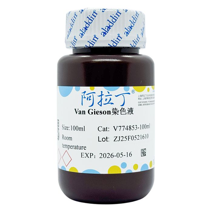
Van Gieson染色液
有货
BioReagent,生物染色剂,用于显微镜 级 ,适用于对基线干扰要求严格的色谱和分析工作流程。
避光,室温。常规运输 。请查阅批次 COA 获取详细规格。
SDS、COA、产品数据表及规格说明书均可下载。可通过批号查询获取批次 COA。
在色谱分析、有机合成和交叉偶联反应领域已被 0 篇同行评审文献引用。
胶原纤维(Collagen Fiber)是结缔组织中分布最广含量最多的一种纤维,广泛分布于各种脏器,其中皮肤、巩膜、肌腱最丰富。Ⅰ型胶原纤维主要是骨、皮肤、肌腱纤维;Ⅱ型胶原纤维主要是软骨胶原;Ⅲ型胶原纤维主要在胚胎组织、成人血管、胃肠道;Ⅳ型胶原纤维主要在基膜中。Van Gieson胶原纤维染色法的染色原理与阴离子染料分子的大小和组织的渗透有关。分子的大小由分子量来体现,小分子量易穿透结构致密、渗透性低的组织,而大分子量则只能进入结构疏松的、渗透性高的组织。PA分子量小,丽春红和复红次之,淡绿分子量最大。VG染色后,肌纤维呈黄色,胶原纤维呈红色。
使用说明:
1、切片脱蜡至水。
2、入Van Gieson染色液中染色3min。
3、用无水酒精快速分化。
4、无水乙醇脱水,二甲苯透明,中性树胶封固。
注意事项:
1、上述试剂有刺激性,请小心操作。
2、为了您的安全和健康,请穿实验服并戴一次性手套操作。
染色结果:
| 胶原纤维 | 红色 |
| 肌纤维、细胞质 | 黄色 |
Collagen Fiber is the most widely‑distributed and most abundant fiber type in connective tissues. It is found in a variety of organs, with the highest abundance in the skin, sclera and tendon. Type Ⅰ collagen fibers are mainly present in bone, skin and tendon; Type Ⅱ collagen fibers are predominantly found in cartilage; Type Ⅲ collagen fibers are located chiefly in embryonic tissues, adult blood vessels and the gastrointestinal tract; Type Ⅳ collagen fibers are primarily localized in basement membranes.The staining principle of the Van Gieson collagen‑fiber staining method is related to the molecular size of anionic dyes and tissue permeability. Molecular size is reflected by molecular weight. Low‑molecular‑weight dyes readily penetrate compact tissues with low permeability, whereas high‑molecular‑weight dyes can only access loose tissues with high permeability. Phosphomolybdic acid (PA) has the lowest molecular weight, followed by Ponceau and fuchsin, and Light Green has the largest molecular weight. After Van‑Gieson (VG) staining, muscle fibers appear yellow and collagen fibers appear red.
Instructions for Use:
1. Deparaffinize sections and rehydrate to water.
2. Stain sections in Van Gieson staining solution for 3 min.
3. Rapid differentiate with absolute ethanol.
4. Dehydrate with absolute ethanol, clear with xylene, and mount with neutral balsam.
Precautions:
1. The above reagents are irritant; handle with care.
2. For your safety and health, wear a lab coat and disposable gloves during operation.
Staining Results:
| Collagen fiber | Red |
| Muscle fiber, Cytoplasm | Yellow |
通过匹配包装上的批号来查找并下载产品的 COA,每批产品都进行了严格的验证,您可放心使用!
| 批号(Lot Number) | 证书类型 | 货号 |
|---|---|---|
 ZJ26F1039775 ZJ26F1039775 |
分析证书 | V774853 |
| ZJ26F1039774 |
分析证书 | V774853 |
| ZJ25F0521611 |
分析证书 | V774853 |
| ZJ26F0333047 |
分析证书 | V774853 |
| ZJ26F0332527 |
分析证书 | V774853 |
| ZJ25F0521610 |
分析证书 | V774853 |
我们的级别选择指南涵盖了目录中所有的变体的纯度、稳定剂状态和应用适用性。
查看 BioReagent 级别指南 → 查看 Biological Stain 级别指南 → 查看 for Microscopy 级别指南 →

 危险品化学品经营许可证(带存储)