L-乳酸由脑、红细胞、骨骼肌和肠产生,由肝脏、心脏和肾脏代谢。人体产生的乳酸几乎全部为L-乳酸。L-乳酸主要由肝脏从血液中清除,缺氧、酸中毒、供氧和低灌注会损害肝脏对L-乳酸的摄取。血液中的L-乳酸浓度对于重症监护和手术期间患者状况的诊断至关重要。高浓度的L-乳酸能指示肠梗塞,心脏骤停和败血症等疾病,唾液中L-乳酸浓度可作为囊性纤维化的早期诊断指标。L-乳酸也是能量代谢,脑和肿瘤细胞的生物标志物。在食品领域,L-乳酸在发酵过程中被用作质量、新鲜度、风味、稳定性、储存寿命的指示剂。
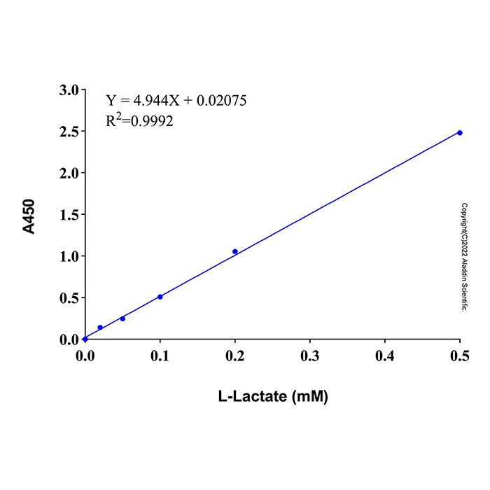
本试剂盒的检测原理为:L-乳酸在L-乳酸脱氢酶(L-Lactate dehydrogenase, L-LDH)的作用下氧化生成丙酮酸(Pyruvate),在这一过程中NAD+还原为NADH;生成的NADH在电子耦合试剂1-mPMS (1-Methoxy-5-methylphenazinium methyl sulfate)的作用下将WST-8还原成橙黄色的Formazan,在450nm左右有最大吸收峰。反应体系中生成的Formazan与样品中L-乳酸含量呈正比。在样品体积为50 μL时可检测含量低至20 µM的L-乳酸,在0.02 mM-0.5 mM (1 nmol-25 nmol)浓度范围内有良好的线性关系。
使用方法
1. 样本制备
(1)血液样品的准备:对于血清样品,将全血在常温下静置30分钟-2小时,4ºC约1000-2000×g离心10分钟,取上清;对于血浆样品,将全血用肝素或者EDTA进行抗凝,4ºC约1000-2000×g离心10分钟,取上清。
(2)细胞样本的准备:对于培养的贴壁细胞,PBS洗涤细胞一次。对于培养的悬浮细胞,离心收集细胞用PBS洗涤细胞一次。按照每100万细胞加入100-200 μL Lysis Buffer的比例加入裂解液,适当吹打,冰浴5-10分钟充分裂解细胞。4ºC, 12,000×g离心5分钟,取上清用于后续检测。
(3)对于组织样品,按照每10 mg组织加入100 μL Lysis Buffer的比例,使用匀浆器冰浴匀浆。4ºC约12,000×g离心5分钟,取上清用于后续检测。
(4)细胞培养上清样品的准备:对于贴壁细胞,直接取培养液;对于悬浮细胞,离心取培养液。
以上所有操作均需在4ºC或冰上操作。制备好的细胞或组织样品如果不能立即检测,可以-20ºC或-80ºC冻存。
2. L-乳酸标准曲线的设置
取2 μL L-Lactate Standard (100 mM),加入398 μL L-Lactate Lysis Buffer或者L-Lactate Assay Buffer混匀(如果是细胞或组织裂解样品,使用L-Lactate Lysis Buffer;如果是血液等其它样品,使用L-Lactate Assay Buffer ),配制成400 μL浓度为0.5 mM L-Lactate Standard。分别取0.5 mM的L-Lactate Standard 0、2、5、10、20、50 μL加入96孔板的标准品孔中,并用对应的Lysis Buffer补足至50 μL,此时,标准曲线的各孔L-乳酸浓度和物质的量分别为0、0.02、0.05、0.1、0.2、0.5 mM和0、1、2.5、5、10、25 nmol。
3. WST-8 Working Solution的配制
按照下表配置WST-8工作液,注意冰上操作。
Samples | 1 | 10 | 20 | 50 | L-Lactate Assay Buffer | 40 μL | 400 μL | 800 μL | 2000 μL | Enzyme Solution | 2 μL | 20 μL | 40 μL | 100 μL | Coenzyme | 4 μL | 40 μL | 80 μL | 200 μL | WST-8 | 2 μL | 20 μL | 40 μL | 100 μL | 1-mPMS | 2 μL | 20 μL | 40 μL | 100 μL | Total volume | 50 μL | 500 μL | 1000 μL | 2500 μL |
|
4. 样本测定
(1)取1-50 μL样品或稀释后的样品至96孔板样品孔中,并加入L-Lactate Lysis Buffer补足50 µL。同时设置仅含L-Lactate Lysis Buffer的孔为空白对照。
(2)各孔加入WST-8显色工作液50 μL,混匀。37ºC避光孵育30分钟。如果颜色较浅,可适当延长反应时间至45或者60分钟;如果孔板的颜色较深,也可以缩短孵育时间为15或者20分钟。
(3)反应结束后,使用酶标仪在450 nm测定吸光度。
5. 结果计算
将标准曲线各孔的信号值减去标准品零浓度孔的信号值。建立标准曲线,并计算样品中L-乳酸的浓度(A),样品孔的信号值应减去空白对照孔的信号值,如果样品的背景对照孔信号比较高,样品的信号值需要减去样品背景对照值。
L-乳酸浓度计算公式如下:
C (mM) = A × n
注1:A为根据标准曲线确定的稀释后的样品L-乳酸浓度(mM);n为样品稀释倍数。
注2:计算获得的L-乳酸浓度其中包含了L-乳酸和L-乳酸根的摩尔浓度,也可以理解为包含了L-乳酸和L-乳酸盐的摩尔浓度。上述仅为了表述方便,仅描述为L-乳酸。如有必要,可根据L-乳酸根的分子量89.07计算出样品中L-乳酸根的质量浓度(μg/ml) = C × 0.08907。
注意事项
1. Lysis Buffer和Assay Buffer需要完全解冻并平衡至室温后再使用,否则会影响检测结果。
2. NADH和NADPH以及会影响NADH和NADPH水平的物质等的存在会对检测产生干扰。如果样品含有干扰物质,须同时设置样品的背景对照孔,即配制WST-8显色工作液时Enzyme Solution用Lactate Assay Buffer替代。计算时样品孔的读数值需要减去背景对照孔的读数。
3. 为确保样品数值在标准曲线范围内,建议进行预实验将样品设置多个稀释倍数,以确定样品中L-乳酸的大致浓度,如果数值不在标准曲线范围内,请调整样品的稀释倍数。
4. 请按照实验需求将组分进行分装,避免反复冻融。
L-lactate is produced by the brain, red blood cells, skeletal muscle, and the intestine, and is metabolized by the liver, heart, and kidneys. Almost all lactate produced in the human body is L-lactate. L-lactate is primarily cleared from the blood by the liver; however, hypoxia, acidosis, impaired oxygen supply, and hypoperfusion can compromise hepatic uptake of L-lactate. The concentration of L-lactate in blood is critical for diagnosing patient conditions during intensive care and surgical procedures. Elevated levels of L-lactate can indicate conditions such as intestinal infarction, cardiac arrest, and sepsis, while the concentration of L-lactate in saliva can serve as an early diagnostic marker for cystic fibrosis. L-lactate also acts as a biomarker for energy metabolism, as well as for brain and tumor cells. In the food industry, L-lactate is used as an indicator of quality, freshness, flavor, stability, and shelf life during fermentation processes.
The detection principle of this kit is as follows: L-lactate is oxidized to pyruvate by L-lactate dehydrogenase (L-LDH), during which NAD⁺ is reduced to NADH. The generated NADH then reduces WST-8 to an orange-yellow formazan dye via the electron coupling reagent 1-mPMS (1-Methoxy-5-methylphenazinium methyl sulfate). The formazan produced exhibits a maximum absorption peak around 450 nm. The amount of formazan generated in the reaction system is proportional to the L-lactate concentration in the sample. With a sample volume of 50 μL, the kit can detect L-lactate levels as low as 20 µM and demonstrates a good linear range from 0.02 mM to 0.5 mM (1 nmol to 25 nmol).
Usage Protocol
1. Sample Preparation
1) Preparation of Blood Samples:
For serum samples: Allow whole blood to clot at room temperature for 30 minutes to 2 hours. Centrifuge at approximately 1000-2000 × g for 10 minutes at 4°C and collect the supernatant.
For plasma samples: Collect whole blood using heparin or EDTA as an anticoagulant. Centrifuge at approximately 1000-2000 × g for 10 minutes at 4°C and collect the supernatant.
2) Preparation of Cell Samples:
For cultured adherent cells: Wash the cells once with PBS; For cultured suspension cells: Centrifuge to collect the cells and wash them once with PBS. Add Lysis Buffer at a ratio of 100-200 µL per 1 million cells. Pipette appropriately to mix and incubate on ice for 5-10 minutes for complete lysis. Centrifuge at 12,000 × g for 5 minutes at 4°C. Collect the supernatant for subsequent assays.
3) Preparation of Tissue Samples:
Add Lysis Buffer at a ratio of 100 µL per 10 mg of tissue. Homogenize the tissue using a homogenizer on ice. Centrifuge at approximately 12,000 × g for 5 minutes at 4°C. Collect the supernatant for subsequent assays.
4) Preparation of Cell Culture Supernatant Samples:
For adherent cells: Directly collect the culture medium; For suspension cells: Centrifuge the culture and collect the supernatant.
All the above procedures must be performed at 4°C or on ice. Prepared cell or tissue samples can be stored at -20°C or -80°C if not assayed immediately.
2. Establishment of L-Lactate Standard Curve
Take 2 μL of L‑Lactate Standard (100 mM) and mix it with 398 μL of either L‑Lactate Lysis Buffer or L‑Lactate Assay Buffer (use L‑Lactate Lysis Buffer for cell or tissue lysate samples; use L‑Lactate Assay Buffer for other samples such as blood) to prepare 400 μL of 0.5 mM L‑Lactate Standard. Then, add 0, 2, 5, 10, 20, and 50 μL of the 0.5 mM L-Lactate Standard into the standard wells of a 96-well plate, respectively. Bring the volume in each well to 50 μL by adding the corresponding L-Lactate Lysis Buffer. As a result, the L-lactate concentrations in the standard curve wells are 0, 0.02, 0.05, 0.1, 0.2, and 0.5 mM, corresponding to amounts of 0, 1, 2.5, 5, 10, and 25 nmol, respectively.
3. Preparation of WST-8 Working Solution
Samples | 1 | 10 | 20 | 50 | L-Lactate Assay Buffer | 40 μL | 400 μL | 800 μL | 2000 μL | Enzyme Solution | 2 μL | 20 μL | 40 μL | 100 μL | Coenzyme | 4 μL | 40 μL | 80 μL | 200 μL | WST-8 | 2 μL | 20 μL | 40 μL | 100 μL | 1-mPMS | 2 μL | 20 μL | 40 μL | 100 μL | Total volume | 50 μL | 500 μL | 1000 μL | 2500 μL |
|
4. Sample Assay
1)Add 1–50 μL of sample or diluted sample to the sample wells of a 96-well plate, and supplement with L-Lactate Lysis Buffer to a total volume of 50 µL. A blank control containing only L-Lactate Lysis Buffer should also be included.
2)Add 50 μL of WST-8 chromogenic working solution to each well and mix thoroughly. Incubate the plate at 37 °C in the dark for 30 minutes. If color development is weak, the reaction time may be extended to 45 or 60 minutes; if color intensity is high, the incubation time may be shortened to 15 or 20 minutes.
3)After the reaction, measure the absorbance at 450 nm using a microplate reader.
5. Calculation of Results
Subtract the signal value of the zero-concentration standard well from that of each standard curve well. Generate a standard curve and calculate the L-lactate concentration in the sample (A). The signal value of the sample well should be corrected by subtracting the value of the blank control well. If the sample background signal is high, subtract the sample background control value accordingly.
The formula for L-lactate concentration is as follows: C (mM) = A × n
Note 1: A represents the L-lactate concentration (mM) of the diluted sample as determined from the standard curve; n is the sample dilution factor.
Note 2: The calculated L-lactate concentration includes the molar concentration of both L-lactic acid and L-lactate ions, which may also be interpreted as the total molar concentration of L-lactic acid and L-lactate salt. For simplicity, the term “L-lactate” is used throughout this description. If required, the mass concentration of L-lactate ions (μg/mL) can be calculated using the molecular weight of L-lactate ion (89.07) as follows: L-lactate ion mass concentration (μg/mL) = C × 0.08907
Precautions
1. Lysis Buffer and Assay Buffer must be completely thawed and equilibrated to room temperature before use, as otherwise the detection results may be affected.
2. The presence of NADH, NADPH, or other substances that influence NADH/NADPH levels may interfere with the assay. If the sample contains potential interfering substances, a sample background control well must be set up by replacing the Enzyme Solution in the WST-8 chromogenic working solution with Lactate Assay Buffer. During calculation, subtract the absorbance value of the background control well from that of the sample well.
3. To ensure that sample values fall within the standard curve range, it is recommended to perform a preliminary experiment with multiple dilution factors to estimate the approximate L-lactate concentration in the sample. If values lie outside the standard curve range, adjust the sample dilution factor accordingly.

 ZJ26F1039743


 危险品化学品经营许可证(带存储)