本试剂盒采用高灵敏度的TUNEL(TdT-mediated dUTP Nick End Labeling)法,旨在简单、快速地检测细胞凋亡过程中发生的DNA片段化。对于经过固定和洗涤的细胞或组织,只要经过一步染色反应,洗涤后就可以通过荧光显微镜或流式细胞仪进行悬浮细胞、贴壁细胞的流式凋亡检测。细胞凋亡时,内源性核酸内切酶被激活,将基因组DNA切割成180-200bp的片段,暴露出大量3‘-OH末端。在末端脱氧核糖核苷酸转移酶(TdT)的催化下,荧光素(如Cy3或AF555)标记的dUTP会掺入到这些断裂位点,从而通过荧光显微镜或流式细胞仪检测到凋亡细胞发出的红色荧光。
本试剂盒适用于多种样本类型,包括石蜡切片、冰冻切片、细胞爬片、细胞涂片及悬浮细胞,无需额外使用二抗,大大简化了实验流程。
使用方法
1. 贴壁细胞(爬片/涂片)
1)固定:吸弃培养基,PBS洗2次。加入4%多聚甲醛室温固定15-30分钟。
2)洗涤:PBS漂洗2次。
3)通透:加入0.2% Triton X-100室温孵育5-10分钟。
4)洗涤:PBS漂洗2-3次,用吸水纸吸干样本周围水分(请勿干燥)。
2. 悬浮细胞
1)收集:收集细胞(约3-5×10⁶个),PBS洗涤1次。
2)固定:加入1-4%多聚甲醛,4°C固定15-30分钟(建议摇床缓慢摇晃以防细胞聚集)。
3)洗涤:离心(300g,5min),弃上清,PBS重悬洗涤1次。
4)通透:加入预冷的70%乙醇(或0.2% Triton X-100),-20°C或冰上孵育30分钟。
5)洗涤:离心,PBS洗涤2次,最后用PBS重悬至约1×10⁶ cells/mL。
3. 石蜡切片
1)脱蜡水化:二甲苯浸泡2次,每次10min,然后用无水乙醇2次,每次5min,95%,85%,75%梯度乙醇各洗涤1次,每次3min,最后用PBS浸泡5min。
2)通透:滴加Proteinase K工作液(20 μg/mL,用PBS稀释),37°C孵育15-30分钟。
3)洗涤:PBS漂洗3次。
4. 冰冻切片
1)复温固定:切片恢复室温,浸入4%多聚甲醛室温固定15-30分钟。
2)洗涤:PBS漂洗2次。
3)通透:滴加Proteinase K工作液,室温孵育10-20分钟。
4)洗涤:PBS漂洗3次。
注意:预处理后的样本建议放在湿盒中保持湿润,防止干片导致高背景。
5. 样本平衡
加入50 μL反应液,轻轻吹打混匀,将样本至于37°C 培养箱平衡15min。
6. TUNEL检测液配制及标记
1)参考以下比例配制工作液,配制时须在冰上避光操作并充分混合均匀,每个样本(50 μL体系)所需体积计算:
反应组分 | 每孔体积(μL) |
TdT酶 | 1 |
荧光标记液 | 2 |
反应缓冲液(5×) | 10 |
去离子水 | 37 |
总体积 | 50 |
2)阴性对照:用ddH₂O替代TdT Enzyme;阳性对照:样本预处理后,加入DNase I工作液(0.3 U/μL)室温孵育10分钟,PBS洗净后再进行TUNEL标记。
3)标记反应:吸尽样本表面的多余液体。滴加 50 μL TUNEL工作液,确保完全覆盖样本。将载玻片置于湿盒中,37°C 避光孵育 60分钟。洗涤:PBS漂洗3次,每次5分钟。
7. 复染、封片与观察
1)核复染:滴加DAPI(1 μg/mL),室温避光孵育5分钟。
2)洗涤:PBS漂洗3次。
3)封片:滴加抗荧光淬灭封片剂,盖上盖玻片。
4)镜检:红色荧光(凋亡细胞):Ex/Em = 550 nm / 570 nm (Cy3通道)。蓝色荧光(所有细胞核):Ex/Em = 340 nm / 488 nm (DAPI通道)。
注意事项
1、避光操作:从加入标记液开始,后续所有步骤均需严格避光,以防荧光淬灭。
2、防止干片:在滴加TUNEL反应液后,若样本体积较小,建议使用湿盒或封口膜覆盖,防止试剂蒸发导致假阴性。
3、阳性对照组需隔离:DNase I处理阳性对照时,必须使用单独的染色缸或离心管,避免DNase I残留污染实验组,导致假阳性。
4、蛋白酶K优化:Proteinase K处理时间过长易导致切片脱落或细胞形态破坏,过短则通透不充分。建议预实验摸索最佳时间。
5、自发荧光:若组织(如肝脏、肾脏)或固定液导致背景荧光较强,可在通透后使用自发荧光淬灭剂处理。
6、安全操作:二甲苯、多聚甲醛及TdT酶均为有害物质,操作时请穿实验服,戴手套。
结果展示

图1. 流式细胞术检测Jurkat细胞凋亡。蓝色曲线为未刺激对照组,绿色曲线为5 μM 星形孢菌素(S102392)处理过夜;虚线:未进行TUNEL标记的细胞,实线:经TUNEL试剂盒标记检测荧光的细胞。

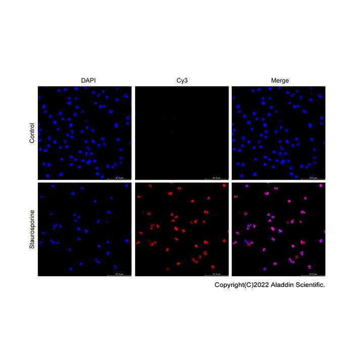
图2. TUNEL荧光染色检测Jurkat细胞凋亡结果。DAPI 蓝色荧光标记全部细胞核,Cy3 红色荧光标记 TUNEL 阳性凋亡细胞;对照组几乎无红色凋亡荧光,星形孢菌素(Staurosporine)处理组可见大量 Cy3 阳性凋亡细胞。
This kit employs a highly sensitive TUNEL (TdT-mediated dUTP Nick End Labeling) method, designed for simple and rapid detection of DNA fragmentation occurring during apoptosis. For fixed and washed cells or tissues, after just a one-step staining reaction and subsequent washing, apoptotic cells can be detected by fluorescence microscopy or flow cytometry for both suspension and adherent cells. During apoptosis, endogenous endonucleases are activated, cleaving genomic DNA into fragments of 180–200 bp and exposing a large number of 3'-OH ends. Under the catalysis of terminal deoxynucleotidyl transferase (TdT), fluorophore-labeled dUTP (such as Cy3, AF555) is incorporated into these break sites, allowing apoptotic cells to be detected by red fluorescence using a fluorescence microscope or flow cytometer.
This kit is suitable for a wide range of sample types, including paraffin-embedded sections, frozen sections, cell smears, cytospin preparations, and suspension cells. It does not require the use of secondary antibodies, greatly simplifying the experimental procedure.
Protocol
1. Adherent cells (coverslip culture / smear preparation)
Fixation: Aspirate culture medium and rinse twice with PBS. Add 4% paraformaldehyde and incubate at room temperature for 15-30 min for fixation.
Washing: Rinse twice with PBS.
Permeabilization: Add 0.3% Triton X-100 and incubate at room temperature for 5–10 min.
Washing: Rinse 2-3 times with PBS; blot residual liquid around specimens with filter paper (avoid complete desiccation of samples).
2. Suspension cells
Cell collection: Harvest approximately 3-5×10⁶ cells and wash once with PBS.
Fixation: Add 1–4% paraformaldehyde, fix at 4 °C for 15-30 min (gentle shaking on a rocker is recommended to prevent cell clumping).
Washing: Centrifuge at 300 g for 5 min, discard supernatant, resuspend pellet in PBS for one round of washing.
Permeabilization: Add pre-cooled 70% ethanol or 0.2% Triton X-100, incubate at −20 °C or on ice for 30 min.
Washing: Centrifuge and wash twice with PBS; finally resuspend cells in PBS to a concentration of ~1×10⁶ cells/mL.
3. Paraffin-embedded tissue sections
Deparaffinization and hydration: Immerse sections in xylene twice (10 min per immersion); sequentially dehydrate in absolute ethanol (2 × 5 min), 95% ethanol, 85% ethanol and 75% ethanol (3 min for each concentration); equilibrate in PBS for 5 min at last.
Permeabilization: Drop Proteinase K working solution (20 μg/mL, diluted with PBS) onto sections and incubate at 37 °C for 15-30 min.
Washing: Rinse three times with PBS.
1. Frozen tissue sections
Rewarming & fixation: Leave sections to equilibrate to ambient temperature, fix in 4% paraformaldehyde at room temperature for 15-30 min.
Washing: Rinse twice with PBS.
Permeabilization: Apply Proteinase K working solution and incubate at room temperature for 10-20 min.
Washing: Rinse three times with PBS.
5、Sample equilibration
Add 50 μL reaction solution, mix gently by pipetting, and equilibrate the samples in a 37 °C incubator for 15 min.
6、Preparation of TUNEL Reaction Mix and Labeling
1)Prepare working solution according to the ratio below. Carry out preparation on ice under dark conditions and mix thoroughly. Calculate required volume per sample (50 μL final system):
Reaction Component | Volume per well (μL) |
TdT Enzyme | 1 |
Fluorescent Labeling Solution | 2 |
Reaction Buffer(5×) | 10 |
Deionized Water | 37 |
Total Volume | 50 |
2)Negative control: Replace TdT Enzyme with ddH₂O . Positive control: After sample pretreatment, incubate specimens with DNase I working solution (0.3 U/μL) at room temperature for 10 min. Thoroughly rinse with PBS prior to TUNEL labeling.
3)Labeling reaction: Aspirate excess liquid on sample surface. Add 50 μL TUNEL working solution to fully cover the specimen. Place slides in a humidified chamber and incubate at 37 °C in the dark for 60 min. Washing step: Rinse 3 times with PBS, 5 min per wash.
7、Counterstaining, Mounting and Microscopic Observation
1)Nuclear counterstaining: Add DAPI (1 μg/mL) solution, incubate at room temperature in the dark for 5 min.
2)Washing: Rinse three times with PBS.
3)Mounting: Drop anti-fade mounting medium and place coverslips.
4)Microscopic observation: Red fluorescence (apoptotic cells): Ex/Em = 550 nm / 570 nm (Cy3 channel); Blue fluorescence (all cell nuclei): Ex/Em = 340 nm / 488 nm (DAPI channel).
Notes
1. Protect from light: From the step of adding the labeling mix onward, all subsequent steps must be strictly performed in the dark (avoid light exposure) to prevent fluorescence quenching.
2. Prevent drying of samples: After adding the TUNEL reaction solution, if the sample volume is small, it is recommended to use a humidified chamber or cover with Parafilm to prevent evaporation of the reagent, which may lead to false-negative results.
3. Isolate positive controls: When treating positive controls with DNase I, a separate staining jar or centrifuge tube must be used to avoid DNase I carryover contaminating the experimental samples, which may cause false-positive results.
4. Optimize Proteinase K treatment: Excessive Proteinase K treatment time can easily lead to section detachment or disruption of cell morphology, while insufficient treatment results in inadequate permeabilization. It is recommended to perform preliminary experiments to determine the optimal incubation time.
5. Autofluorescence: If tissues (e.g., liver, kidney) or the fixative solution cause strong background fluorescence, treat the samples with an autofluorescence quencher after permeabilization.
6. Safety precautions: Xylene, paraformaldehyde, and TdT enzyme are all hazardous substances. Please wear a lab coat and gloves during handling.
Results Presentation
Figure 1. Flow cytometric analysis of apoptosis in Jurkat cells. Blue curve represents the untreated control group, and green curve corresponds to cells treated overnight with 5 μM staurosporine (Cat# S102392). Dashed lines: cells without TUNEL labeling; solid lines: cells stained with TUNEL kit for fluorescence detection.
Figure 2. Detection of apoptosis in Jurkat cells via TUNEL fluorescence staining. All cell nuclei were counterstained with DAPI (blue), and TUNEL-positive apoptotic cells were labeled with Cy3 (red). Few red apoptotic signals were observed in the control group, whereas abundant Cy3-positive apoptotic cells were detected in the staurosporine-treated group.

 ZJ26F0737054


 危险品化学品经营许可证(带存储)