显色法TUNEL细胞凋亡检测试剂盒(Colorimetric TUNEL Apoptosis Assay Kit)为您提供了一种高灵敏度又快速简便的细胞凋亡检测方法。对于待检测的细胞或组织样品,经过生物素标记和后续的DAB显色等步骤,即可在普通光学显微镜下观察到凋亡细胞。
细胞凋亡是程序性细胞死亡的一种形式。根据程序性细胞死亡过程中的细胞变化,细胞凋亡通常分为早期、中期和晚期。细胞凋亡后期的特征是核形态、染色质凝集、核膜降解和DNA断裂的变化。
TUNEL (TdT-mediated dUTP Nick-End Labeling)法检测细胞凋亡的原理为:细胞在发生凋亡时,会激活一些DNA内切酶,剪断核小体间的基因组DNA,而正常的或正在增殖的细胞中几乎没有DNA的断裂,因而没有3'-OH形成,很少能够被标记。暴露的3'-OH可在末端脱氧核酸转移酶(TdT)的催化下加上生物素标记的dUTP,辣根过氧化物酶(HRP)标记的链霉亲和素(Streptavidin-HRP)可与之结合,在HRP的催化下通过DAB显色可检测到凋亡细胞,从而实现细胞凋亡检测。
产品组分
T1456509 | 组分 | 物理外观 | 20 T | 50 T | 储存条件 | T1456509A | TdT Enzyme | 液体 | 20 μL | 50 μL | -20℃. | T1456509B | Biotin-11-dUTP | 液体 | 40 μL | 100 μL | -20℃. | T1456509C | 反应缓冲液(5×) | 液体 | 200 μL | 500 μL | -20℃. | T1456509D | Streptavidin-HRP | 液体 | 20 μL | 50 μL | -20℃. 避光保存. | T1456509E | DAB Solution A | 液体 | 1 mL | 2.5 mL | -20℃. 避光保存. | T1456509F | DAB Solution B | 液体 | 1 mL | 2.5 mL | -20℃. 避光保存. | T1456509G | DNase I (10U/μL) | 液体 | 10 μL | 25 μL | -20℃. | T1456509H | DNase I 稀释液 | 液体 | 1 mL | 2.5 mL | -20℃. |
注:本试剂盒20T包装可以检测20个涂片、切片或96孔板、48孔板、24孔板或12孔板的样品;可以检测10个6孔板的样品;本试剂盒50T包装可以检测50个涂片、切片或96孔板、48孔板、24孔板或12孔板的样品;可以检测25个6孔板的样品。 |
使用方法
实验材料(自备)
1. 1×PBS (pH 7.4)
2. 0.1% Triton X-100(1×PBS配制)
3. 4%多聚甲醛(1×PBS配制)
4. 3% H₂O₂
5. ddH2O
6. 苏木素
实验步骤
1. 样本处理
A. 悬浮细胞或细胞悬液
a) 细胞收集:400 g离心5分钟收集细胞(不超过200万细胞)。PBS洗涤2次。
b) 固定:加入适量4%多聚甲醛重悬细胞,4℃固定30分钟。400 g离心5分钟收集细胞,PBS洗涤2次。
c) 通透:加入适量0.1% Triton X 100,室温通透15分钟。400 g离心5分钟收集细胞,PBS洗涤2次。
d) 封闭:每孔样本加入100 μL 3% H₂O₂ 覆盖细胞,室温避光孵育20分钟以灭活内源性过氧化氢酶。PBS洗涤2次。
B. 贴壁细胞或细胞爬片
a) 洗涤:用PBS洗涤细胞一次。
注:可以干燥样品使细胞贴的更牢固。
b) 固定:加入适量4%多聚甲醛,室温固定30分钟。PBS洗涤2次。
c) 通透:加入适量0.1% Triton X 100,室温通透15分钟。PBS洗涤2次。
d) 封闭:每孔样本加入100 μL 3% H2O2覆盖细胞,室温避光孵育20分钟以灭活内源性过氧化氢酶。PBS洗涤2次。
C. 石蜡组织切片
a) 石蜡组织切片脱蜡与水化:二甲苯中脱蜡5-10分钟。换用新鲜的二甲苯,再脱蜡5-10分钟。无水乙醇5分钟。90%乙醇2分钟。70%乙醇2分钟,蒸馏水2分钟。
b) 通透:将蛋白酶K (2 mg/mL)用PBS稀释至终浓度为20 μg/mL,每个样本滴加100 μL覆盖样本,37℃孵育15-30分钟。
注:孵育时间过长会增加组织切片在后续洗涤步骤中从载玻片上脱落的风险,过短则可能造成透性处理不充分,影响标记效率;不同组织的最佳作用温度和时间需自行摸索。
c) 洗涤:PBS洗涤3次,每次5分钟。
注:这一步必须把蛋白酶K洗涤干净,否则会严重干扰后续的标记反应。
d) 封闭:加入适量3% H₂O₂ ,室温孵育20分钟以灭活内源性过氧化氢酶。
e) 洗涤:PBS洗涤2次,每次5分钟。用滤纸吸去多余液体,保持样品湿润。
D. 冰冻组织切片
a) 固定:将切片样本浸入4%多聚甲醛中,室温固定30分钟。PBS缓慢漂洗3次,每次5分钟。
b) 通透:将蛋白酶K (2 mg/mL)用PBS稀释至终浓度为20 μg/mL,每个样本滴加100 μL覆盖样本,37℃孵育10分钟。
注:孵育时间过长会增加组织切片在后续洗涤步骤中从载玻片上脱落的风险,过短则可能造成透性处理不充分,影响标记效率;不同组织的最佳作用温度和时间需自行摸索。
c) PBS漂洗切片3次,每次5分钟。
d) 封闭:将切片样本浸入3% H₂O₂ 溶液,室温孵育20分钟,以灭活切片内源的过氧化氢酶。
e) PBS漂洗切片3次,每次5分钟,用滤纸吸干切片样本周围液体,用免疫组化笔圈好样本轮廓,以便下游标记。将处理好的切片放在湿盒中保持湿润。
注:若在后续实验操作中发现免疫组化笔画的轮廓圈被破坏,需及时补画。
E. 阳性对照(选做)
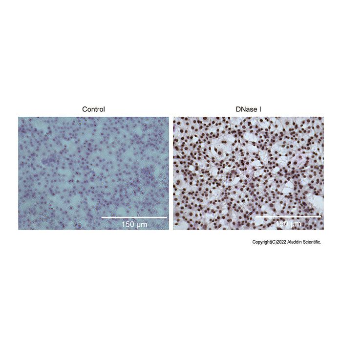
a) 将DNase I dilution solution (10×)用ddH₂O稀释至1×备用。
b) 将DNase I (10 U/μL)用DNase I dilution solution (1×)稀释至终浓度为20 U/mL。
c) 加入100 μL DNase I dilution solution (1×)到已处理的样品上,覆盖样品并室温平衡5分钟。
d) 弃掉DNase I dilution solution (1×),加入100 μL DNase I (20 U/mL),室温孵育10-30分钟。
e) 弃掉DNase I,PBS洗涤2次。
2. TUNEL反应液(现配现用)
TUNEL反应液组分 | 1个样品 | 5个样品 | 10个样品 | TdT Enzyme | 1 μL | 5 μL | 10 μL | Biotin-11-dUTP | 2 μL | 10 μL | 20 μL | 反应缓冲液(5×) | 10 μL | 50 μL | 100 μL | ddH2O | 37 μL | 185 μL | 370 μL | 总体积 | 50 μL | 250 μL | 500 μL |
注:生物素TUNEL反应缓冲液和TdT Enzyme需完全溶解混匀后使用。 |
3. 样品生物素标记
A. 每个样品加入50 μL TUNEL反应液,使反应液覆盖样品。37℃孵育1小时。
注:50 μL生物素标记液适合涂片、切片或96孔板、48孔板、24孔板或12孔板的一个孔,如果是6孔板中的一个孔生物素标记液宜使用100 μL。孵育时注意防护防止工作液挥发。
B. 弃掉TUNEL反应液,用PBS (含0.1% TritonX-100 和5 mg/mL BSA)洗涤3次。
4. 显色反应
A. Streptavidin-HRP工作液的配制
Streptavidin-HRP工作液 | 1个样品 | 5个样品 | 10个样品 | Streptavidin-HRP | 1 μL | 5 μL | 10 μL | PBS | 99 μL | 495 μL | 990 μL | 总体积 | 100 μL | 500 μL | 1000 μL |
注:配制好的Streptavidin-HRP工作液必须一次使用完毕,不宜冻存。 |
A. DAB显色液的配制:按照每个样品使用0.2-0.5 mL显色液的比例配制适量DAB显色液。等体积混合适量DAB Solution A和DAB Solution B,充分混匀后即为DAB显色液。
注:配制好的DAB显色液必须一次使用完毕,不宜冻存。
B. 反应:在样品上加100 μL Streptavidin-HRP工作液,室温孵育30分钟。
C. 洗涤:弃掉Streptavidin-HRP工作液,用PBS洗涤2次。
D. 显色:滴加0.2-0.5 mL DAB显色液,室温孵育5-30分钟或根据显色情况孵育适当时间。
注:如果显色很强可以短于5分钟即停止显色,如果显色很弱,可以适当延长显色时间。
A. 终止:弃掉DAB显色液,PBS洗涤3次以终止显色反应。
B. 细胞核染色(可选):用苏木素染色液或甲基绿染色液进行细胞核染色。
C. 洗涤:弃掉细胞核染色液,PBS洗涤2次。
D. 直接进行观察,或用95%乙醇脱水5分钟,再用100%乙醇脱水2次,每次约3分钟,再用二甲苯透明2次,每次5分钟,随后封片观察。
注意事项
1. 使用前请将产品瞬时离心至管底,再进行后续实验。
2. 本产品仅限于专业人员的科学研究用,不得用于临床诊断或治疗,不得用于食品或药品。
3. 为了您的安全和健康,请穿实验服并戴一次性手套操作。
4. 叠氮化钠 (NaN3) 会抑制 HRP,因此反应体系中不能含有叠氮化钠。
5. 荧光标记液和TdT Enzyme避免反复冻融,建议不要涡旋。
6. 实验过程中请保持样本的湿润,防止干片造成的实验失败。
The Colorimetric TUNEL Apoptosis Assay Kit provides a highly sensitive, rapid, and straightforward method for detecting apoptotic cells. Following biotin-labeling and subsequent DAB development, apoptotic cells can be visualized under a standard light microscope.
Apoptosis is a form of programmed cell death, typically categorized into early, mid, and late stages based on characteristic cellular changes. Late-stage apoptosis is marked by alterations in nuclear morphology, including chromatin condensation, nuclear membrane degradation, and DNA fragmentation.
The principle of the TUNEL (TdT-mediated dUTP Nick-End Labeling) method for detecting cell apoptosis is as follows: When cells undergo apoptosis, some DNA endonucleases are activated, which cut the genomic DNA between nucleosomes. In normal or proliferating cells, there is almost no DNA breakage, so no 3'-OH is formed and few can be labeled. The exposed 3'-OH can be labeled with biotin-labeled dUTP under the catalysis of terminal deoxynucleotidyl transferase (TdT). Streptavidin-HRP labeled chains can bind to it. Through the catalysis of HRP, it can be detected by DAB color development, thus enabling the detection of cell apoptosis.
Kit Contents
T1456509 | Components | Appearance | 20 T | 50 T | Storage | T1456509A | TdT Enzyme | Liquid | 20 μL | 50 μL | -20℃. | T1456509B | Biotin-11-dUTP | Liquid | 40 μL | 100 μL | -20℃. | T1456509C | Reaction Buffer(5×) | Liquid | 200 μL | 500 μL | -20℃. | T1456509D | Streptavidin-HRP | Liquid | 20 μL | 50 μL | -20℃. Store in the dark. | T1456509E | DAB Solution A | Liquid | 1 mL | 12.5 mL | -20℃. Store in the dark. | T1456509F | DAB Solution B | Liquid | 1 mL | 12.5 mL | -20℃. Store in the dark. | T1456509G | DNase I (10U/μL) | Liquid | 10 μL | 25 μL | -20℃. | T1456509H | DNase I dilution solution | Liquid | 1 mL | 12.5 mL | -20℃. |
Note: The 20T packaging of this kit can test 20 slides, sections or 96-well plates, 48-well plates, 24-well plates or 12-well plates of samples; it can test 10 6-well plates of samples; the 50T packaging of this kit can test 50 slides, sections or 96-well plates, 48-well plates, 24-well plates or 12-well plates of samples; it can test 25 6-well plates of samples. |
Usage method
Materials to Be Supplied by the User
1. 1X PBS (pH 7.4)
2. 0.1% Triton X-100 (Prepared in 1X PBS)
3. 4% Paraformaldehyde (Prepared in 1X PBS)
4. 3% H₂O₂
5. ddH₂O
6. Hematoxylin
Experimental Procedure
1. Sample Preparation
A. Suspension Cells or Cell Suspensions
a) Cell Collection: Centrifuge at 400 g for 5 minutes to collect the cells (no more than 2 million cells). Wash twice with PBS.
b) Fixation: Resuspend cells in an adequate volume of 4% Paraformaldehyde and fix at 4°C for 30 min. Centrifuge at 400 g for 5 min. Wash twice with PBS.
c) Permeabilization: Resuspend cells in an adequate volume of 0.1% Triton X-100 and incubate at room temperature (RT) for 15 min. Centrifuge at 400 g for 5 min. Wash twice with PBS.
d) Blocking: Add 100 μL of 3% H₂O₂ to cover the cell pellet. Incubate at RT in the dark for 20 min. Wash twice with PBS.
B. Adherent Cells or Cell Climbing Slides
a) Wash: Wash the cells once with PBS.
b) Fixation: Add an adequate volume of 4% Paraformaldehyde and fix at RT for 30 min. Wash twice with PBS.
c) Permeabilization: Add an adequate volume of 0.1% Triton X-100 and incubate at RT for 15 min. Wash twice with PBS.
d) Endogenous Peroxidase Blocking: Add 100 μL of 3% H₂O₂ to cover the cells. Incubate at RT in the dark for 20 min. Wash twice with PBS.
C. Paraffin-Embedded Tissue Sections
a) Deparaffinization and Hydration: Incubate in xylene for 5-10 min. Repeat with fresh xylene. Then incubate sequentially in: 100% ethanol (5 min), 90% ethanol (2 min), 70% ethanol (2 min), and finally distilled water (2 min).
b) Permeabilization: Dilute Protease K (2 mg/mL) to 20 μg/mL in PBS. Apply 100 μL to cover the tissue section and incubate at 37°C for 15-30 min.
c) Wash: Wash the section three times with PBS, 5 min each.
d) Blocking: Apply an adequate volume of 3% H₂O₂ and incubate at RT for 20 min.
e) Wash: Wash the section twice with PBS, 5 min each. Carefully remove excess liquid, ensuring the sample remains hydrated.
D. Frozen Tissue Sections
a) Fixation: Immerse sections in 4% Paraformaldehyde and fix at RT for 30 min. Rinse gently three times with PBS, 5 min each.
b) Permeabilization: Dilute Protease K (2 mg/mL) to 20 μg/mL in PBS. Apply 100 μL to cover the tissue section and incubate at 37°C for 10 min.
c) Wash: Rinse sections three times with PBS, 5 min each.
d) Blocking: Immerse sections in 3% H₂O₂ and incubate at RT for 20 min.
e) Wash: Rinse sections three times with PBS, 5 min each. Carefully dry around the section and use a hydrophobic pen to circle the tissue.
E. Positive Control (Optional)
a) Dilute the 10× DNase I Dilution Solution to 1× using ddH₂O.
b) Dilute DNase I (10 U/μL) to 20 U/mL using the 1X DNase I Dilution Solution.
c) Apply 100 μL of 1X DNase I Dilution Solution to the prepared sample and incubate at RT for 5 min for equilibration.
d) Remove the solution, apply 100 μL of diluted DNase I (20 U/mL), and incubate at RT for 10 - 30 min.
e) Remove DNase I solution and wash the sample twice with PBS.
2. TUNEL Reaction Mixture (Prepare Fresh)
Components of the TUNEL reaction solution | One sample | Five samples | Ten samples | TdT Enzyme | 1 μL | 5 μL | 10 μL | Biotin-11-dUTP | 2 μL | 10 μL | 20 μL | Reaction Buffer(5×) | 10 μL | 50 μL | 100 μL | ddH₂O | 37 μL | 185 μL | 370 μL | Total Volume | 50 μL | 250 μL | 500 μL |
Note: The biotin TUNEL reaction buffer and TdT Enzyme should be thoroughly dissolved and mixed before use. |
3. Biotin Labeling of Samples
A. Apply 50 μL of TUNEL Reaction Mixture to each sample, ensuring complete coverage. Incubate at 37°C for 60 min in a humidified chamber.
B. Remove the TUNEL Reaction Mixture. Wash three times with PBS (containing 0.1% Triton X-100 and 5 mg/mL BSA).
4. Color Development
A. Preparation of Streptavidin-HRP Working Solution:
Streptavidin-HRP working solution | One sample | Five samples | Ten samples | Streptavidin-HRP | 1 μL | 5 μL | 10 μL | PBS | 995 μL | 495 μL | 990 μL | Total Volume | 100 μL | 500 μL | 1000 μL |
Note: The prepared Streptavidin-HRP working solution must be used up completely at one time and should not be frozen. |
B. Preparation of DAB Coloring Solution
Prepare an appropriate amount of DAB color-developing solution by using 0.2 - 0.5 mL of the color-developing solution for each sample. Mix equal volumes of DAB Solution A and DAB Solution B, and after thorough mixing, it will be the DAB color-developing solution.
Note: The prepared DAB color-developing solution must be used up completely at one time and should not be frozen for storage.
C. Reaction: Add 100 μL of Streptavidin-HRP working solution onto the sample and incubate at room temperature for 30 minutes.
D. Washing: Discard the Streptavidin-HRP working solution and wash twice with PBS.
E. Color development: Add 0.2 - 0.5 mL of DAB color development solution and incubate at room temperature for 5 - 30 minutes or for an appropriate time depending on the color development.
Note: If the color development is strong, stop the color development within 5 minutes. If the color development is weak, extend the incubation time appropriately.
F. Termination: Discard the DAB color development solution and wash three times with PBS to terminate the color development reaction.
G. Cell nucleus staining (optional): Use hematoxylin staining solution or methyl green staining solution for cell nucleus staining.
H. Washing: Discard the cell nucleus staining solution and wash twice with PBS.
I. Observe directly, or dehydrate with 95% ethanol for 5 minutes, then dehydrate twice with 100% ethanol for about 3 minutes each time, then clear with xylene twice for 5 minutes each time, and then mount for observation.
Matters needing attention
1. Briefly centrifuge all vials prior to opening to bring the contents to the bottom.
2. For research use only. Not for use in diagnostic procedures.
3. Wear lab coats and disposable gloves for safety.
4. Sodium azide (NaN₃) is an inhibitor of HRP and must not be present in any solution.
5. Avoid repeated freeze-thaw cycles of the Biotin-11-dUTP and TdT Enzyme. Vortexing is not recommended.
6. Keep samples hydrated throughout the procedure to prevent drying artifacts.

 ZJ26F0737039


 危险品化学品经营许可证(带存储)