
计算溶液所需的质量、体积或浓度。
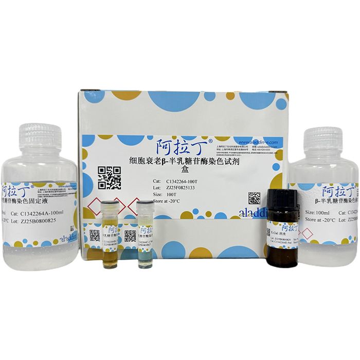
细胞衰老β-半乳糖苷酶染色试剂盒
有货
BioReagent,用于显微镜 级 ,适用于对基线干扰要求严格的色谱和分析工作流程。
避光,-20°C储存,避免反复冻融。超低温运输 。请查阅批次 COA 获取详细规格。
SDS、COA、产品数据表及规格说明书均可下载。可通过批号查询获取批次 COA。
在色谱分析、有机合成和交叉偶联反应领域已被 0 篇同行评审文献引用。
细胞衰老(Cell senescence)是指细胞在执行生命活动过程中,随着时间的推移,细胞增殖与分化能力和生理功能逐渐发生衰退的变化过程。通常情况下,机体会对衰老细胞进行清除,而未被清除的衰老细胞则可能会体内积累,引起低水平的炎症,或进一步诱导临近组织衰退、癌变等。细胞衰老的一些特征包括细胞形态改变、细胞周期停滞、衰老相关分泌表型、大分子损伤及代谢紊乱等。目前,鉴别细胞衰老特异性最强的标志为衰老相关 β-半乳糖苷酶 (senescence associated βgalactosidase, SA-β-Gal),随着细胞的衰老其会逐渐累积,而在衰老前细胞、静止细胞和终末分化细胞中则是缺乏的。
本试剂盒利用衰老细胞中的β-Gal在pH为6.0时可以催化5-溴-4-氯-3-吲哚基-β-D-吡喃半乳糖苷(5-bromo-4-chloro-3-indolyl-β-D-galactopyranoside, X-gal)水解生成蓝色物质,从而可在光学显微镜下观察到细胞或组织的衰老状况。
产品组分表
|
储存条件
-20℃保存,其中 E 组分 X-Gal 溶液需避光保存。
以贴壁细胞(6孔板)举例,每孔1 mL染色工作液,100 mL可用于100个孔的染色。
使用方法
1. 贴壁细胞染色(6 孔板)
(1)细胞铺板过夜,待细胞生长至 60% - 80%汇合度时可以进行实验。
(2)去除细胞培养液,用 PBS 清洗 1 次。
(3)加入 1 mL β-半乳糖苷酶染色固定液,室温固定 10~15 min。
(4)去除 β-半乳糖苷酶染色固定液,用 PBS 清洗 3 次,每次 2-3 min。
(5)根据表1配置染色工作液,具体染色工作液总量请根据样本类型、样本数等进行计算。
表1. 染色工作液
|
注:① 为减少样本染色过程中出现结晶现象,建议X-Gal使用前可适当37℃加热1-3 h。② 染色工作液需要现配现用,并尽量在15 min内用完。
(6)去除PBS,每孔加入1 mL染色工作液,37℃无CO2培养箱孵育过夜。
注:为防止液体蒸发影响染色结果,建议在边缘孔中加入水或PBS,减少“边缘效应”,同时用封口膜或保鲜膜封住孔板。
(7)普通光学显微镜下进行观察计数。
(8)可选:去除染色工作液,加入 2 mL PBS,4℃可保存数天。
2. 悬浮细胞染色
(1)收集细胞,2500 ×g 离心 10 min,用 PBS 清洗 1 次。
(2)加入 1 mL β-半乳糖苷酶染色固定液,室温固定 10-15 min。
注:可将细胞放置摇床上缓慢摇动,以避免细胞结团。
(3)2500 ×g 离心 10 min,去除 β-半乳糖苷酶染色固定液,用 PBS 清洗 3 次,每次 1-3 min。
(4)根据表 1 配置染色工作液,具体染色工作液总量请根据样本类型、样本数等进行计算。
(5)2500 ×g 离心 10 min,去除 PBS。每管加入 0.5-1 mL 染色工作液,37℃孵育过夜。
(6)吸取部分细胞滴加到载玻片上或 6 孔板内,普通光学显微镜下进行观察计数。
(7)可选:去除染色工作液,再加入 1 mL PBS,4℃可保存数天。
注意事项
1. 使用前请将产品瞬时离心至管底,再进行后续实验。
2. 细胞衰老 β-半乳糖苷酶检测依赖于特定的 pH 条件,而 CO2 培养箱中较高浓度的 CO2会影响染色工作液的 pH 值,因此,37℃孵育不能在 CO2培养箱中进行。
3. X-Gal溶液在 -20℃或4℃保存会冻结,室温或37℃水浴2-5 min并适当摇动即可完全融解,若需要分装该溶液或配置染色工作液,需要使用聚丙烯 (PP) 或玻璃材质的耗材,不要使用聚苯乙烯(PS)耗材,如细胞培养板、血清移液管等。
4. β-半乳糖苷酶染色液B的管底可能存在少量沉淀,属正常现象,请充分震荡涡旋,待全部溶解后再进行染色实验。
5. 如需染色切片样本,请参考相关文献并进行预实验摸索方案。
6. β-半乳糖苷酶染色固定液存在一定的毒性和腐蚀性,操作时请穿实验服并戴一次性口罩和手套,避免直接接触人体或吸入体内。
Cell senescence refers to a progressive degenerative process in cells. Over time, their capacity for proliferation and differentiation, as well as physiological functions, gradually decline during vital activities. Normally, senescent cells are cleared by the organism. Unremoved senescent cells may accumulate in the body, triggering low-grade inflammation, and further inducing deterioration of adjacent tissues or even carcinogenesis. Typical characteristics of cell senescence include altered cell morphology, cell cycle arrest, senescence-associated secretory phenotype, macromolecular damage and metabolic disorders. At present, senescence-associated β-galactosidase (SA-β-Gal) is the most specific biomarker for identifying cell senescence. Its level gradually rises as cells senesce, while it is undetectable in pre-senescent cells, quiescent cells and terminally differentiated cells.
This kit takes advantage of the property that β-galactosidase in senescent cells can catalyze the hydrolysis of 5-bromo-4-chloro-3-indolyl-β-D-galactopyranoside (X-gal) at pH 6.0 to produce blue precipitates. The senescent status of cells or tissues can therefore be observed under a light microscope.
Product Components
| C1342264 | Component | 100T | Storage |
| C1342264A | β-Galactosidase Staining Fixative | 100 mL | -20℃. .Avoid freeze/thaw cycle. |
| C1342264B | β-Galactosidase Staining Solution A | 1.5 mL | 20℃. Avoid freeze/thaw cycle. |
| C1342264C | β-Galactosidase Staining Solution B | 1.5 mL | -20℃. Avoid freeze/thaw cycle. |
| C1342264D | β-Galactosidase Staining Solution C | 100 mL | -20℃. Avoid freeze/thaw cycle. |
| C1342264E | X-Gal Solution | 5 mL | -20℃. Avoid freeze/thaw cycle. Store in the dark. |
Storage Conditions
Store all components at -20 °C. Component E (X-Gal Solution) requires protection from light.
Taking adherent cells cultured in 6-well plates as an example, add 1 mL of staining working solution per well. A total volume of 100 mL is sufficient for 100 wells.
Instructions for Use
1. Staining Protocol for Adherent Cells (6-well Plate)
①Seed cells and incubate overnight. Perform the experiment when cell confluence reaches 60%–80%.
②Discard the culture medium and rinse cells once with PBS.
③Add 1 mL of β-galactosidase staining fixative, and incubate at room temperature for 10–15 minutes for fixation.
④Remove the fixative, then rinse with PBS three times, 2–3 minutes each time.
⑤Prepare the staining working solution according to Table 1. Calculate the total volume based on sample type and quantity.
Table 1. Preparation of Staining Working Solution
Components | Preparation for 1 mL working solution |
β-Galactosidase Staining Solution A | 10 µL |
β-Galactosidase Staining Solution B | 10 µL |
β-Galactosidase Staining Solution C | 940 µL |
X-Gal Solution | 40 µL |
Notes: To reduce crystal formation during staining, warm X-Gal solution at 37 °C for 1–3 hours before use. Prepare the staining working solution freshly before use and consume it within 15 minutes.
⑥Aspirate PBS, add 1 mL of staining working solution to each well, and incubate overnight at 37 °C in a CO₂-free incubator.
Note: To avoid staining artifacts caused by liquid evaporation, fill the peripheral wells with water or PBS to minimize the edge effect. Seal the plate with parafilm or plastic wrap.
⑦Observe and count cells under a conventional light microscope.
⑧Optional step: Remove the staining working solution, add 2 mL of PBS. Samples can be stored at 4 °C for several days.
2. Staining of Suspension Cells
①Harvest cells and centrifuge at 2500 ×g for 10 minutes, then rinse once with PBS.
②Add 1 mL of β-galactosidase staining fixative and fix cells at room temperature for 10–15 minutes.
Note: Gently shake the cells on a shaker to prevent cell clumping.
③Centrifuge at 2500 ×g for 10 minutes and discard the fixative. Rinse cells with PBS three times, 1–3 minutes each time.
④Prepare the staining working solution according to Table 1. Calculate the required total volume based on sample type and quantity.
⑤Centrifuge at 2500 ×g for 10 minutes and remove PBS. Add 0.5–1 mL of staining working solution to each tube and incubate overnight at 37 °C.
⑥Pipette an aliquot of cell suspension onto a glass slide or into a 6-well plate, then observe and count cells under a standard light microscope.
⑦Optional: Remove the staining working solution and add 1 mL of PBS. Samples can be stored at 4 °C for several days.
Precautions
①Briefly centrifuge all reagents to spin down liquid to the bottom of tubes before use.
②Senescence-associated β-galactosidase staining requires a specific pH environment. High CO₂ concentration in a CO₂ incubator will alter the pH of the staining working solution. Therefore, incubation at 37 °C must be performed in a CO₂-free incubator.
③X-Gal solution will freeze at -20 °C or 4 °C. Thaw completely by water bathing at room temperature or 37 °C for 2–5 minutes with gentle shaking. Use polypropylene (PP) or glassware for aliquoting X-Gal solution or preparing working solution. Do not use polystyrene (PS) consumables such as cell culture plates and serological pipettes.
④A small amount of precipitate may form at the bottom of the tube containing β-galactosidase Staining Solution B, which is normal. Vortex thoroughly until fully dissolved prior to staining.
⑤For tissue section staining, refer to relevant literature and optimize the protocol through preliminary experiments.
⑥The β-galactosidase staining fixative is toxic and corrosive. Wear a lab coat, disposable mask and gloves during operation. Avoid direct skin contact and inhalation.
通过匹配包装上的批号来查找并下载产品的 COA,每批产品都进行了严格的验证,您可放心使用!
| 批号(Lot Number) | 证书类型 | 货号 |
|---|---|---|
 ZJ26F0636751 |
分析证书 | C1342264 |
| ZJ25F0825133 |
分析证书 | C1342264 |


 危险品化学品经营许可证(带存储)