背景介绍:
乳酸脱氢酶细胞毒性检测试剂盒(WST-8法) 是一种基于WST-8的显色反应,通过比色法检测细胞毒性时释放的乳酸脱氢酶(Lactate dehydrogenase, LDH)活性或检测其它样品中的LDH活性的试剂盒。
LDH催化反应生成NADH,后者与WST-8反应产生在450nm处有特征吸收的水溶性甲臜染料,通过测定吸光度即可定量LDH活性,吸光度值与酶活性呈正比。
该试剂盒以LDH释放作为细胞膜完整性的指标,为细胞毒性检测提供了一种高灵敏度、操作安全的非放射性方法。相较于传统INT法,WST-8法具有灵敏度更高、吸光度变化更明显、结果更准确的特点。
使用说明:
1. 试剂盒的准备工作:
LDH检测工作液(LDH assay working solution)的配制:按照每个检测反应100µl的体积配制适量的LDH检测工作液。均匀混合91μl检测缓冲液(Detection assay buffer)和9μl显色液(Chromogen solution),即可配制成100µl LDH检测工作液。根据待检测样品(包括对照)的数量,配制适量的LDH检测工作液。需现配现用,配制和使用过程中均要注意适当避光。
2. LDH标准曲线的制作(选做):
如果希望进行LDH酶活性的绝对定量,需自备LDH标准品,并新鲜配制不同浓度LDH标准品,如0、65、125、250、500、1000mU/ml等。如有必要,在后续实验中可以根据样品的LDH活性,对标准曲线的浓度范围进行适当调整。
3. 检测细胞数的确定(选做):
通常按照常规的细胞接种量进行接种即可。但由于每种细胞LDH量有所差异,为了得到最好的显色效果,可以通过预实验确定最适细胞数。
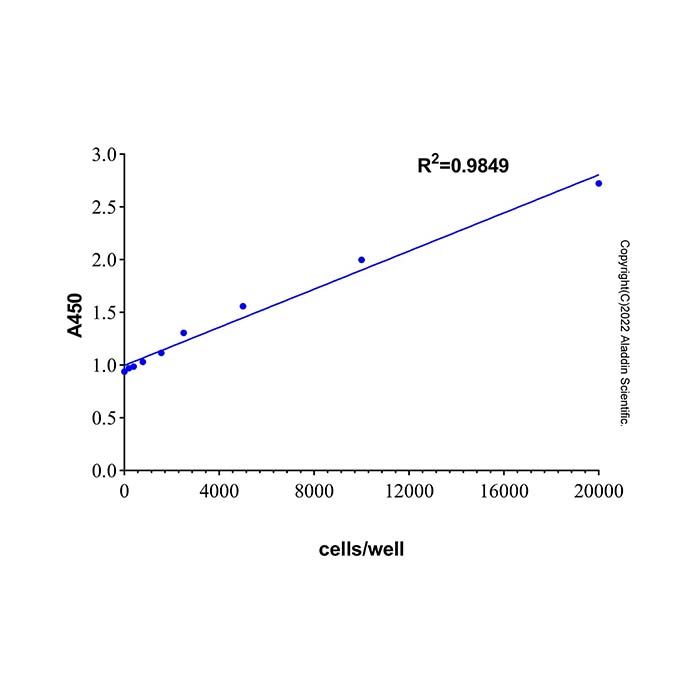
1. 收集细胞,用培养液洗涤细胞后配制成每毫升20万个细胞的细胞悬液,取0、12.5、25、50、100μl细胞悬液接种到96孔细胞培养板中,即每孔细胞数为0、2500、5,000、10,000、20,000个。分两组,一组是总LDH组(Total LDH)、一组是未处理对照组(Untreated control),每个细胞浓度3个重复,过夜培养。
2. 37ºC继续培养一定时间(与正式实验中的药物处理时间一致)。总LDH组各孔加入10μl LDH释放试剂,未处理对照组各孔加入10μl培养液。混匀,室温(约25ºC)避光孵育15分钟。
3. 每孔加入100μl LDH检测工作液。混匀,室温(约25ºC)避光孵育0-30分钟,由于不同细胞差异较大,建议首次实验时,分别测定0、5、10、20、30分钟时的吸光度,以便确定最佳反应时间。此时可用铝箔避光包裹后置于水平摇床或侧摆摇床上缓慢摇动。孵育完毕后在450nm处测定吸光度。如无450nm滤光片,可以使用420-480nm的滤光片。可以选择设定600nm(或600nm以上,如650nm)作为参比波长(也称参考波长)。450nm吸光度的读数减去参比波长的吸光度读数即可作为实测读数(A450)。测试总LDH读数不超过但邻近酶标仪吸光度检测上限的细胞密度及孵育时间,即为最适细胞数和最适测试时间。
4. 细胞毒性样品的准备:
1. 按照下表设置各实验组别,将适量细胞接种到96孔板中,过夜培养。按照下表中各组别处理方法更换培养液。注:为确保实验有效性,建议每个组别设置三个复孔。
Groups | Method | Note |
未处理对照(Untreated control) | 更换100μl的新鲜培养液 | 含细胞 |
背景对照(Background control) | 更换100μl的新鲜培养液 | 不含细胞* |
总LDH(Total LDH) | 更换100μl的新鲜培养液 | 含细胞 |
总LDH背景(Background of total LDH) | 更换100μl的新鲜培养液 | 不含细胞* |
样品(Sample) ** | 更换100μl含药培养液*** | 含细胞 |
样品背景(Background of Sample) | 更换100μl含药培养液*** | 不含细胞* |
阳性对照(Positive Control)(选做,如步骤2) | 更换100μl适当稀释后的LDH标准品*** |
*为了避免培养液中的血清、药物、LDH释放试剂对读数的影响,需设置不含细胞、但加入了相同培养液的组,用于消除背景。
**样品组也可以是仅用于乳酸脱氢酶检测的样品。
***含药培养液:根据实验需要,在新鲜培养液中加入药物(例如加入0-5μl特定的药物)。可设置不同浓度,不同处理时间。
2. 到达药物处理的预定时间后,总LDH组和总LDH背景组加入10μl的LDH释放试剂,其它组加入10μl的培养液。混匀,室温(约25ºC)避光孵育15分钟。
5. 吸光度测定:
1. 如果样品组细胞无其它用途,可以直接在每孔中加入100μl LDH检测工作液。如果样品组细胞还有其它用途,可直接取各孔的上清液100μl,或将培养板用多孔板离心机400×g室温离心5分钟,分别取各孔的上清液100μl。然后加入到一个新的96孔板中,接着加入100μl LDH检测工作液。
2. 混匀,室温(约25ºC)避光孵育10-30分钟,孵育的最佳时间可以通过步骤3的预实验确定。孵育时可用铝箔包裹后置于水平摇床或侧摆摇床上缓慢摇动。
3. 在每个孔中加入50μl终止液,混匀,然后在450nm处测定吸光度。如无450nm滤光片,可以使用420-480nm的滤光片。可以选择设定600nm(或600nm以上,如650nm)作为参比波长(也称参考波长),450nm吸光度的读数扣除参比波长的吸光度读数即可作为实测读数(A450)。
注意:为确保检测效果,可以不添加终止液,在不同时间点测定吸光度,直到获得比较理想的吸光度数据为止,然后再酌情添加终止液。
4. 取各组复孔的平均值计算各组别吸光度。
样品组吸光度 = A450样品 - A450样品背景。
总LDH组吸光度 = A450总LDH- A450总LDH背景。
未处理对照组吸光度 = A450未处理对照 - A450空白对照背景。
细胞毒性或死亡率 = (样品组吸光度 -未处理对照组吸光度) / (总LDH组吸光度 - 未处理对照组吸光度) × 100%
5. 可绘制细胞毒性曲线:纵坐标为样品组吸光度,横坐标为药物浓度;据此可计算该药物作用特定时间的半致死剂量LD50。
6. 若需准确计算出LDH酶活性的绝对活性,可通过一系列LDH标准品及相应测得的吸光度值绘制标准曲线,通过标准曲线相应公式计算出样品中LDH的酶活性。各孔数值减去背景对照后,以检测的吸光度(OD450)为纵坐标,LDH酶活力(mU)为横坐标,绘制LDH标准曲线,同时计算出该趋势线的公式,这样就可以计算出样品中的LDH酶活力了。
A450nm = k × LDH酶活力单位(mU) + b,通过Excel等软件计算出趋势线的斜率k和截距b。
根据上述公式计算样品中LDH活力。
检测体系中样品LDH酶活力单位(mU) = (样品组吸光度 - b) / k
样品中LDH酶活力(mU/ml) = 检测体系中LDH酶活力单位(mU) / 检测样品体积
注意事项:
1. 冷冻会使样品中部分乳酸脱氢酶失活,样品在4ºC可放置2-3天。建议样品准备好后尽量当天完成测定。
2. 在检测细胞培养液中的乳酸脱氢酶时,由于血清含有乳酸脱氢酶,使用含血清的培养液会增加背景读数,在检测时一定要设置没有细胞,但加入了相同体积培养液的对照孔,以用于消除背景。血清含量越高,背景值越高。如果对于实验无明显影响,建议使用灭活血清,这样血清中的乳酸脱氢酶会被很大程度上失活,大幅降低背景。如果对于实验无明显影响,实验时可以使用无血清培养液或血清浓度较低的培养液,这样能有效降低血清中乳酸脱氢酶的本底活性。
3. 细胞过度生长、密度过高、离心速度过大、培养箱内外温差过大,都会造成细胞释放乳酸脱氢酶增加。此外,不同的细胞乳酸脱氢酶的含量也存在一定差异。
4. 未加终止液时,随着时间延长,吸光值会逐渐增大。加入终止液后,显色可以稳定保存。
5. 如果希望进行乳酸脱氢酶活性的绝对定量,用户需自备乳酸脱氢酶标准品。
Background Introduction:
The Lactate Dehydrogenase Cytotoxicity Assay Kit (WST-8 Method) is a kit based on the WST-8 chromogenic reaction, which detects the activity of lactate dehydrogenase (LDH) released during cytotoxicity or LDH activity in other samples through colorimetric analysis.
LDH catalyzes the reaction to generate NADH, which then reacts with WST-8 to produce a water-soluble formazan dye with characteristic absorption at 450 nm. By measuring the absorbance, LDH activity can be quantified, with absorbance values being directly proportional to enzyme activity.
This kit uses LDH release as an indicator of cell membrane integrity, providing a highly sensitive, operationally safe, and non-radioactive method for cytotoxicity detection. Compared with the traditional INT method, the WST-8 method offers higher sensitivity, more pronounced absorbance changes, and more accurate results.
Instruction for use
1. Kit Preparations:
Prepare an appropriate amount of LDH assay working solution based on a volume of 100 µl per detection reaction. For every 100 µl of working solution, uniformly mix 91 µl of Detection Assay Buffer with 9 µl of Chromogen Solution. Prepare a sufficient volume according to the total number of samples to be tested (including controls).
Note: The working solution should be prepared immediately before use. Protect it from light during both preparation and use.
2. Preparation of the LDH Standard Curve (Optional) :
To achieve absolute quantification of LDH enzyme activity, it is necessary to prepare an LDH standard separately. Freshly prepare LDH standards at different concentrations, such as 0, 65, 125, 250, 500, and 1000 mU/ml. If necessary, the concentration range of the standard curve can be appropriately adjusted in subsequent experiments based on the LDH activity of the samples.
3. Determination of Cell Number for Assay (Optional) :
Typically, cells can be seeded according to the routine seeding density. However, since the LDH content varies among different cell types, it is recommended to determine the optimal cell number through preliminary experiments to achieve the best chromogenic results.
1. Harvest the cells, wash them with culture medium, and prepare a cell suspension at a density of 200,000 cells per milliliter. Aliquot 0, 12.5, 25, 50, and 100 µl of the cell suspension into a 96-well cell culture plate, corresponding to 0, 2,500, 5,000, 10,000, and 20,000 cells per well, respectively. Set up two groups: a Total LDH group and an Untreated Control group, with three replicates for each cell concentration. Incubate the plate overnight.
2. Continue incubation at 37°C for a specified period (consistent with the drug treatment duration in the formal experiment). Add 10 µl of LDH Release Reagent to each well of the Total LDH group, and add 10 µl of culture medium to each well of the Untreated Control group. Mix well and incubate at room temperature (approximately 25°C) protected from light for 15 minutes.
3. Add 100 µl of LDH assay working solution to each well. Mix well and incubate at room temperature (approximately 25°C) protected from light for 0–30 minutes. Due to significant variations among different cell types, it is recommended that during the initial experiment, absorbance readings be taken at 0, 5, 10, 20, and 30 minutes to determine the optimal reaction time. During incubation, the plate may be wrapped in aluminum foil and placed on a horizontal or orbital shaker with gentle agitation. After incubation, measure the absorbance at 450 nm. If a 450 nm filter is not available, a filter within the range of 420–480 nm may be used. A reference wavelength of 600 nm (or above, e.g., 650 nm) can be optionally set. The measured absorbance at 450 nm (A450) is obtained by subtracting the absorbance at the reference wavelength from the reading at 450 nm. The cell density and incubation time that yield a total LDH reading approaching but not exceeding the upper detection limit of the microplate reader are considered the optimal cell number and test time, respectively.
4. Preparation of Cytotoxicity Samples:
1. Set up the experimental groups as described in the table below. Seed an appropriate number of cells into a 96-well plate and culture overnight. Then, replace the culture medium according to the treatments specified for each group in the table.
Note: To ensure experimental validity, it is recommended to set up three replicate wells for each group.
Groups | Method | Note |
Untreated control | Replace with 100 µl of fresh culture medium | Containing cells |
Background control | Replace with 100 µl of fresh culture medium | Cell-free * |
Total LDH | Replace with 100 µl of fresh culture medium | Containing cells |
Background of total LDH | Replace with 100 µl of fresh culture medium | Cell-free * |
Sample ** | Replace with 100 µl of drug-containing culture medium *** | Containing cells |
Background of Sample | Replace with 100 µl of drug-containing culture medium *** | Cell-free * |
Positive Control(Optional) | Replace with 100 µl of appropriately diluted LDH standard *** |
*To avoid interference from serum, drugs, or LDH release reagent present in the culture medium on the readings, it is necessary to include a cell‑free group treated with the same culture medium for background subtraction.
** The sample group may also refer to samples intended solely for lactate dehydrogenase detection.
*** Drug‑containing culture medium: According to experimental requirements, add the drug (e.g., 0–5 µl of a specific drug) to fresh culture medium. Different concentrations and different treatment durations can be set.
2. Upon reaching the predetermined duration of drug treatment, add 10 µl of LDH Release Reagent to the Total LDH group and the Total LDH Background group. For all other groups, add 10 µl of culture medium. Mix well and incubate at room temperature (approximately 25°C) protected from light for 15 minutes.
5. Absorbance Measurement:
1. If the cells in the sample wells are not required for any other purposes, 100 µl of LDH assay working solution can be added directly to each well. If the cells in the sample wells are needed for other uses, either directly collect 100 µl of supernatant from each well, or centrifuge the culture plate at 400 × g for 5 minutes at room temperature using a plate centrifuge, then collect 100 µl of supernatant from each well. Transfer the supernatant to a new 96-well plate, and then add 100 µl of LDH assay working solution to each well.
2. Mix well, and incubate at room temperature (approximately 25°C) protected from light for 10–30 minutes. The optimal incubation time can be determined based on the preliminary experiment described in Step 3. During incubation, the plate may be wrapped in aluminum foil and gently shaken on a horizontal or orbital shaker.
3. Add 50 µl of stop solution to each well, mix well, and then measure the absorbance at 450 nm. If a 450 nm filter is not available, a filter within the range of 420–480 nm may be used. A reference wavelength of 600 nm (or above, e.g., 650 nm) can be optionally set. The measured absorbance value (A450) is obtained by subtracting the absorbance at the reference wavelength from the absorbance reading at 450 nm.
Note: To ensure optimal detection, the stop solution may be omitted initially. The absorbance can be measured at different time points until relatively ideal absorbance data are obtained, after which the stop solution may be added as appropriate.
4. Calculate the average absorbance for each group from the replicate wells.
Sample group absorbance = A450 sample − A450 sample background.
Total LDH group absorbance = A450 Total LDH − A450 Total LDH background.
Untreated control group absorbance = A450 untreated control − A450 blank control background.
Cytotoxicity or mortality = (Sample group absorbance − Untreated control group absorbance) / (Total LDH group absorbance − Untreated control group absorbance) × 100%
5. A cytotoxicity curve can be plotted with the absorbance of the sample group on the y‑axis and the drug concentration on the x‑axis. From this curve, the half‑lethal dose (LD₅₀) of the drug at a specific treatment time can be calculated.
6. To accurately calculate the absolute activity of LDH enzyme, a standard curve can be established using a series of LDH standards and their corresponding measured absorbance values. The enzyme activity of LDH in the sample can then be determined via the formula derived from the standard curve.
After subtracting the background control value from each well, plot the LDH standard curve with the measured absorbance (OD₄₅₀) on the y‑axis and LDH enzyme activity (mU) on the x‑axis. The formula of the resulting trendline should also be calculated, which allows for the determination of LDH enzyme activity in the sample.
A₄₅₀nm = k × LDH enzyme activity (mU) + b
Use software such as Excel to calculate the slope (k) and intercept (b) of the trendline.
Based on the above formula, calculate the LDH activity in the sample:
LDH enzyme activity in the assay system (mU) = (Absorbance of the sample group – b) / k
LDH enzyme activity in the sample (mU/ml) = LDH enzyme activity in the assay system (mU) / volume of sample tested
Matters needing attention
1. Freezing may partially inactivate lactate dehydrogenase in the samples. The samples can be stored at 4°C for 2–3 days. It is recommended to complete the measurement on the same day the samples are prepared whenever possible.
2. When detecting lactate dehydrogenase in cell culture medium, it should be noted that serum contains LDH. Using serum‑containing medium will increase the background reading. Therefore, it is essential to include control wells without cells but containing the same volume of culture medium for background subtraction. The higher the serum concentration, the higher the background value. If it does not significantly affect the experiment, it is recommended to use inactivated serum, which can largely deactivate the LDH present in serum and substantially reduce the background. Alternatively, if experimental conditions allow, serum‑free or low‑serum culture medium can be used during the assay to effectively lower the background activity of LDH from serum.
3. Overgrowth of cells, high cell density, excessive centrifugation speed, and large temperature differences inside and outside the incubator can all lead to increased LDH release from cells. Additionally, the LDH content varies to some extent among different cell types.
4. In the absence of stop solution, the absorbance will gradually increase over time. After adding the stop solution, the color development can remain stable.
5. If absolute quantification of lactate dehydrogenase activity is desired, users need to prepare their own LDH standard separately.

 ZJ26F0433840


 危险品化学品经营许可证(带存储)