乳酸脱氢酶(lactate dehydrogenase,LDH或LD)是一种稳定的蛋白质,存在于正常细胞的胞质中,正常情况下不能透过细胞膜;当细胞受损伤时,膜通透性增强,LDH即被释放到胞外;细胞质内LDH减少,培养液中LDH增多,测定培养液中LDH活性或LDH漏出率即可反映药物的细胞毒性。LDH属于氧化还原酶,能可逆的催化乳酸(L)和丙酮酸(P)之间的氧化还原反应。其反应公式:乳酸+NAD+→丙酮酸+NADH+H⁺,其中:L→P为正向反应;P→L为逆向反应。
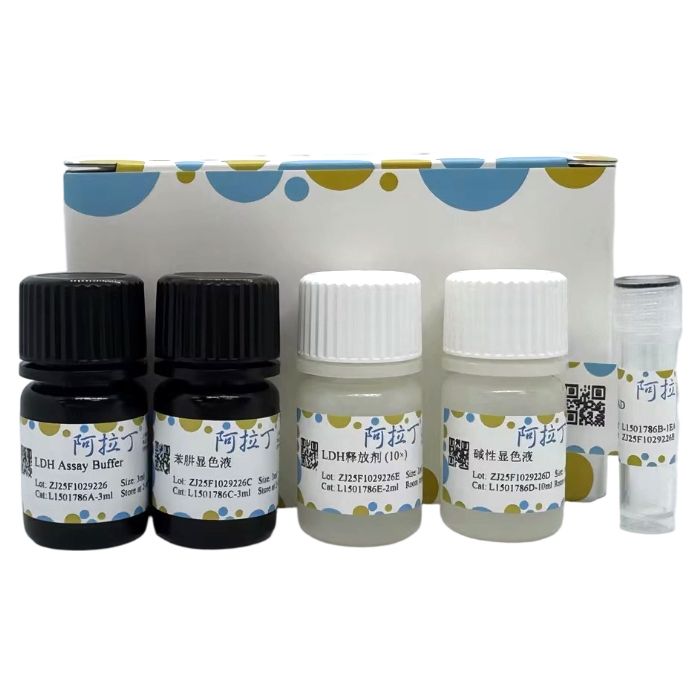
检测原理:以NAD为受氢体,LDH催化乳酸脱氢生成丙酮酸,丙酮酸再与二硝基苯肼反应生成丙酮酸二硝基苯腙,后者在碱性溶液中呈棕红色,颜色深浅与丙酮酸浓度呈正比,可用酶标仪检测440nm处的吸光度,通过公式可以计算出细胞毒性时释放的LDH活性或检测其他样品中的LDH活性,可用于常规的LDH活性的检测,更常用于以LDH释放为指标的细胞毒性的检测。该试剂盒仅用于科研领域,不适用于临床诊断或其他用途。
L1501786
| Component
| 100T
| 500T
| Storage
|
L1501786A
| LDH Assay Buffer | 3 mL | 15 mL
| 2-8℃. Store in the dark.
|
L1501786B
| NAD | 1EA
| 2EA
| -20℃
|
L1501786C
| 苯肼显色液 | 3 mL | 15 mL | 2-8℃. Store in the dark.
|
L1501786D
| 碱性显色液 | 10 mL | 50 mL | RT. |
L1501786E
| LDH释放剂 (10×)
| 2 mL | 10 mL | RT. |
自备仪器和试剂
1、96孔板培养的待测组和对照组细胞样品、无菌PBS、培养液、蒸馏水
2、多孔板离心机、96孔板或离心机、离心管、恒温箱或水浴锅、酶标仪
实验流程
1、样品准备
1.1 LDH释放检测
根据细胞的大小和生长速度将适量细胞接种到96培养板中,使待检测时细胞密度不超过90%满,吸去培养液,PBS清洗一次,加新培养液,并根据实验需要设置相应的背景空白对照孔A(无细胞的培养液)、样品对照孔B(未经药物处理的对照细胞)、最大酶活对照孔C(未经药物处理的细胞后经裂解的样品)、药物处理样品孔D(经药物处理的细胞)等分组,继续培养;待检测前取出细胞培养板,在“最大酶活对照孔C”加入LDH释放剂 (10×),加入量为原有培养液体积的10%,并反复吹打混匀数次,然后继续培养1h左右;将细胞培养板用多孔板离心机400g离心5min,分别抽取各孔上清液5μl,加入到一个新的96孔板相应孔中,用于后续的LDH检测。
1.2 细胞内总LDH的细胞毒性和细胞增殖检测
根据细胞的大小和生长速度将适量细胞接种到96培养板中,使待检测时细胞密度不超过90%满。加入不同药物处理,并设置适当对照,将细胞培养板用多孔板离心机400g离心5min,吸除培养液,加入150μl用PBS 10倍稀释的LDH释放剂,晃动培养板使充分混匀,然后继续培养1h左右;将细胞培养板用多孔板离心机400g离心5min,分别抽取各孔上清液5μl,加入到一个新的96孔板相应孔中,用于后续的细胞毒性检测。
1.3 蛋白浓度测定
样品准备完毕后可以用BCA蛋白浓度测定试剂盒测定蛋白浓度(推荐使用阿拉丁B665595 BCA蛋白定量试剂盒或R1491648 即用型BCA蛋白定量试剂盒),以便于后续计算单位蛋白重量组织或细胞内的LDH含量。
2、配制NAD溶液
取1支NAD(粉末),溶解于1.5ml去离子水,即成。
3、LDH 酶促
按照下表顺序依次加入各溶液,并注意避免产生气泡;如果样品中酶活性过高,可减少样品用量或适当稀释后再行测定。
| 试剂(μL) | 加入量(μL) |
| 待测样品(上清液) | 5 |
| LDH Assay Buffer | 25 |
| NAD溶液 | 5 |
混匀,37℃孵育15min。
混匀,37℃孵育15min。
4、LDH测定
混匀,室温放置5min,酶标仪440nm处测定各孔吸光度。
5、结果计算
细胞毒性或死亡率=(A D - A B )/(A C - A B )x100%
如同时检测一已知浓度c的LDH酶标准品对应吸光度值A γ 和标准空白对照吸光度值A γ0 ,则可粗略计算出样品中的酶活力:
待测样品中LDH活力单位 (mU/ml) =(A B -A A )/(A γ -A γ0 )xc
如需准确计算样品LDH酶的绝对活性,可用自备的LDH标准品及测得的相应吸光度值绘制标准曲线,通过标准曲线相应公式可计算出样品的酶活性。
其中:
A A =背景空白对照孔A的吸光度
A B =样品对照孔B的吸光度
A C =最大酶活对照孔C的吸光度
A D =药物处理样品孔D的吸光度
6、结果与分析
通过直接比较每孔LDH的活性判定药物或毒物的细胞毒性,LDH的活性越高,表示细胞膜通透性越高,细胞受损越严重。
注意事项
1、培养细胞时要用无血清或低浓度血清的培养液,以排除血清的干扰,否则会有偏差。
2、EDTA对LDH有抑制作用,操作中避免使用或尽量彻底清除含EDTA的试剂。
3、LDH尽可能现取现测,如收集的细胞培养液放置时间较长,可能使LDH的活性降低。
4、同一批试验尽量用同一次配置的溶液,溶液的使用量应统一,反应时间也应一致。
5、酶促反应中,上清液取样量以 2.5~10μL为宜,如果样品中酶活性过高,可减少样品用量或适当稀释后再行测定。
6、显色后应在15min内测定完成。
7、碱性显色液有一定腐蚀性,请小心操作。
8、试剂开封后请尽快使用,以防影响后续实验效果。
9、为了您的安全和健康,请穿实验服并戴一次性手套操作。
Lactate dehydrogenase (LDH or LD) is a stable protein present in the cytoplasm of normal cells and normally cannot pass through the cell membrane. When cells are damaged, membrane permeability increases, and LDH is released extracellularly. A decrease in intracellular LDH and an increase in LDH in the culture medium occur. Measuring the LDH activity in the culture medium or the LDH leakage rate can reflect drug-induced cytotoxicity. LDH belongs to the oxidoreductase family and can reversibly catalyze the redox reaction between lactate (L) and pyruvate (P). The reaction formula is: Lactate + NAD⁺ → Pyruvate + NADH + H⁺, where L → P is the forward reaction and P → L is the reverse reaction.
Detection Principle: Using NAD⁺ as a hydrogen acceptor, LDH catalyzes the dehydrogenation of lactate to generate pyruvate. Pyruvate then reacts with dinitrophenylhydrazine to form pyruvate dinitrophenylhydrazone, which appears brownish-red in an alkaline solution. The color intensity is proportional to the pyruvate concentration. The absorbance at 440 nm can be measured using a microplate reader. The released LDH activity during cytotoxicity or the LDH activity in other samples can be calculated using formulas. This kit can be used for routine LDH activity detection and is more commonly used for cytotoxicity assays using LDH release as an indicator.
This kit is for scientific research use only and is not intended for clinical diagnosis or other purposes.
L1501786
| Component
| 100T
| 500T
| Storage
|
L1501786A
| LDH Assay Buffer | 3 mL | 15 mL
| 2-8℃. Store in the dark.
|
L1501786B
| NAD | 1EA
| 2EA
| -20℃
|
L1501786C
| Phenylhydrazine Color Solution | 3 mL | 15 mL | 2-8℃. Store in the dark.
|
L1501786D
| Alkaline Color Solution | 10 mL | 50 mL | RT. |
L1501786E
| LDH Releasing Agent (10X) | 2 mL | 10 mL | RT. |
User-Prepared Instruments and Reagents
1. 96-well plate cultured test and control group cell samples, sterile PBS, culture medium, distilled water.
2. Microplate centrifuge, 96-well plate or centrifuge, centrifuge tubes, incubator or water bath, microplate reader.
Experimental Procedure
1. Sample Preparation
1.1 LDH Release Assay
Seed an appropriate number of cells into a 96-well culture plate based on cell size and growth rate, so that the cell density does not exceed 90% confluency at the time of detection.
Aspirate the culture medium, wash once with PBS, add fresh culture medium.
Set up corresponding control groups according to experimental needs:
Background Blank Control Well A: Culture medium without cells.
Sample Control Well B: Control cells without drug treatment.
Maximum Enzyme Activity Control Well C: Lysed samples from untreated cells.
Drug-treated Sample Well D: Cells treated with the drug.
Continue cultivation.
Before detection, take out the cell culture plate. Add LDH Releasing Agent (10X) to the "Maximum Enzyme Activity Control Well C" at a volume equal to 10% of the original culture medium volume. Mix thoroughly by pipetting up and down several times. Continue cultivation for about 1 hour.
Centrifuge the cell culture plate at 400 g for 5 minutes using a microplate centrifuge.
Aspirate 5 µL of supernatant from each well and transfer it to the corresponding wells of a new 96-well plate for subsequent LDH detection.
1.2 Cytotoxicity and Cell Proliferation Assay for Intracellular Total LDH
Seed an appropriate number of cells into a 96-well culture plate based on cell size and growth rate, so that the cell density does not exceed 90% confluency at the time of detection.
Treat with different drugs and set up appropriate controls.
Centrifuge the cell culture plate at 400 g for 5 minutes using a microplate centrifuge.
Aspirate the culture medium.
Add 150 µL of LDH Releasing Agent diluted 10-fold with PBS. Shake the plate to mix thoroughly. Continue cultivation for about 1 hour.
Centrifuge the cell culture plate at 400 g for 5 minutes using a microplate centrifuge.
Aspirate 5 µL of supernatant from each well and transfer it to the corresponding wells of a new 96-well plate for subsequent cytotoxicity detection.
1.3 Protein Concentration Determination
After sample preparation, the protein concentration can be determined using a BCA Protein Assay Kit (Aladdin B665595 BCA Protein Quantification Kit or R1491648 Ready-to-Use BCA Protein Quantification Kit are recommended) to facilitate subsequent calculation of LDH content per unit protein weight in tissues or cells.
2. Preparation of NAD Solution
Take one vial of NAD (powder) and dissolve it in 1.5 mL of deionized water.
3. LDH Enzymatic Reaction
Add solutions sequentially according to the table below, taking care to avoid bubbles. If the enzyme activity in the sample is too high, reduce the sample volume or dilute appropriately before assay.
Reagent (µL)
| Volume (µL)
|
Test Sample (supernatant)
| 5 |
LDH Assay Buffer
| 25 |
NAD Solution
| 5 |
Mix well, incubate at 37°C for 15 min.
Phenylhydrazine Color Solution
| 25 |
Mix well, incubate at 37°C for 15 min.
| Alkaline Color Solution | 100 |
| Distilled Water | 150 |
4. LDH Measurement
Mix well and let stand at room temperature for 5 minutes. Measure the absorbance of each well at 440 nm using a microplate reader.
5. Result Calculation
Cytotoxicity or Mortality Rate (%) = (A D - A B ) / (A C - A B ) × 100%
If the absorbance value A γ of a known concentration *c* of an LDH enzyme standard and the absorbance value A γ0 of the standard blank control are measured simultaneously, the enzyme activity in the sample can be roughly calculated:
LDH Activity in Test Sample (mU/mL) = (A B - A A ) / (A γ - A γ0 ) × *c*
For accurate calculation of the absolute LDH enzyme activity in the sample, use a self-prepared LDH standard to plot a standard curve with the measured absorbance values. The enzyme activity of the sample can be calculated using the formula derived from the standard curve.
Where:
A A = Absorbance of Background Blank Control Well A
A B = Absorbance of Sample Control Well B
A C = Absorbance of Maximum Enzyme Activity Control Well C
A D = Absorbance of Drug-treated Sample Well D
6. Results and Analysis
The cytotoxicity of drugs or toxicants can be determined by directly comparing the LDH activity in each well. Higher LDH activity indicates higher cell membrane permeability and more severe cell damage.
Precautions
1. Use serum-free or low-serum concentration culture medium when culturing cells to exclude serum interference; otherwise, deviations may occur.
2. EDTA inhibits LDH. Avoid using or thoroughly remove reagents containing EDTA during operation.
3. Measure LDH as soon as possible after collection. If the collected cell culture medium is stored for too long, LDH activity may decrease.
4. Use solutions prepared at the same time for the same batch of experiments. The volume of solutions used and the reaction time should be consistent.
5. In the enzymatic reaction, the recommended supernatant sample volume is 2.5-10 µL. If the enzyme activity in the sample is too high, reduce the sample volume or dilute appropriately before assay.
6. Measurement should be completed within 15 minutes after color development.
7. The Alkaline Color Solution is somewhat corrosive; handle with care.
8. Use reagents promptly after opening to avoid affecting subsequent experimental results.
9. For your safety and health, please wear a lab coat and disposable gloves during operation.

 ZJ25F1029226


 危险品化学品经营许可证(带存储)