Fibroblast growth factors (FGFs) comprise a family of at least eighteen structurally related proteins that are involved in a multitude of physiological and pathological cellular processes, including cell growth, differentiation, angiogenesis, wound healing and tumorgenesis. The biological activities of the FGFs are mediated by a family of type I transmembrane tyrosine kinases which undergo dimerization and autophosphorylation after ligand binding. Four distinct genes encoding closely related FGF receptors, FGF R1-4, are known. All four genes for FGF Rs encode proteins with an N-terminal signal peptide, three immunoglobulin (Ig)-like domains, an acid‑box region containing a run of acidic residues between the IgI and IgII domains, a transmembrane domain and the split tyrosine-kinase domain. Multiple forms of FGF R1‑3 are generated by alternative splicing of the mRNAs. A frequent splicing event involving FGF R1 and 2 results in receptors containing all three Ig domains, referred to as the alpha isoform, or only IgII and IgIII, referred to as the beta isoform. Only the alpha isoform has been identified for FGF R3 and FGF R4. Additional splicing events for FGF R1-3, involving the C-terminal half of the IgIII domain encoded by two mutually exclusive alternative exons, generate FGF receptors with alternative IgIII domains (IIIb and IIIc). A IIIa isoform which is a secreted FGF binding protein containing only the N-terminal half of the IgIII domain plus some intron sequences has also been reported for FGF R1. Mutations in FGF R1-3 have been found in patients with birth defects involving craniosynostosis. The complex patterns of expression of these receptors as well as the specificity of their interactions with the various FGF ligand family members are under investigation.
Post-translational: Autophosphorylated. Binding of FGF family members together with heparan sulfate proteoglycan or heparin promotes receptor dimerization and autophosphorylation on tyrosine residues. Autophosphorylation occurs in trans between the two FGFR molecules present in the dimer. Phosphorylation at Tyr-724 is essential for stimulation of cell proliferation and activation of PIK3R1, STAT1 and MAP kinase signaling. Phosphorylation at Tyr-760 is required for interaction with PIK3R1 and PLCG1.Ubiquitinated. Is rapidly ubiquitinated after ligand binding and autophosphorylation, leading to receptor internalization and degradation. Subject to both proteasomal and lysosomal degradation.N-glycosylated in the endoplasmic reticulum. The N-glycan chains undergo further maturation to an Endo H-resistant form in the Golgi apparatus.
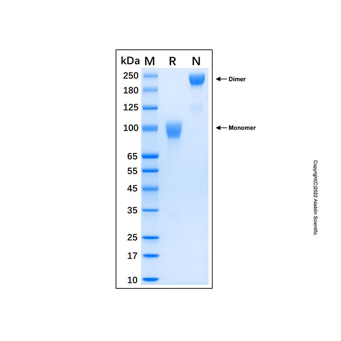
97.0 kDa, under reducing conditions; 226.1 kDa, under non-reducing conditions.
分子类型
蛋白质
产品线
生物产品
储存与运输
物理形态
冻干(Lyophilized)
储存缓冲液
Lyophilized from PBS, pH 7.4
浓度
见COA
复溶
We recommend that this vial be briefly centrifuged prior to opening to bring the contents to the bottom. Reconstitute at 1.0 mg/mL in sterile distilled water. Stock solutions should be apportioned into working aliquots and stored at ≤ -20 °C. Further dilutions should be made in appropriate buffered solutions.
储存条件
-20°C储存,避免反复冻融
运输条件
超低温运输
稳定性与储存
-20℃保存 12个月;复溶后建议分装,避免反复冻融。
图片
Recombinant Human FGFR3 (IIIc) Chimera Protein (rp184034) - SDS-PAGE 3 μg/lane of Recombinant Human FGFR3 (IIIc) Chimera Protein was resolved with SDS-PAGE under reducing (R) and non-reducing (N) conditions and visualized by Coomassie® Blue staining, showing a band at 97.0 kDa under reducing conditions and 226.1 kDa under non-reducing conditions. Immunoglobulins are composed of two identical heavy chains and two identical light chains; disulfide-linked (PubMed:20176268).

 ZJ25F0724936


 危险品化学品经营许可证(带存储)