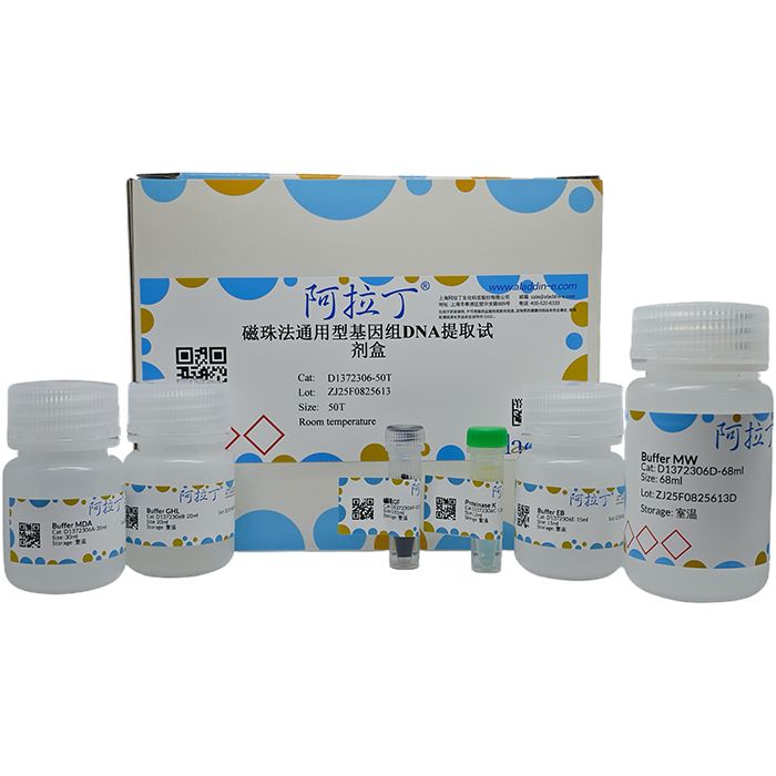
本试剂盒采用超顺磁性的磁性粒子和优化的缓冲液,从血液、干血斑、唾液、口腔拭子、动物组织、细菌、细胞和FFPE样本中分离纯化高质量基因组DNA。磁性粒子在一定pH和盐离子条件下对核酸具有很强且特异的亲和力,而当条件改变时,磁性粒子释放吸附的核酸,能够达到快速分离纯化核酸的目的。整个操作过程安全便捷,提取的基因组DNA片段大,纯度高,质量稳定可靠,尤其适合高通量工作站的自动化提取。使用本试剂盒纯化的DNA适用于各种常规操作,包括酶切、PCR、荧光定量PCR、文库构建、Southern杂交、芯片检测和高通量测序等实验。
自备试剂
RNase A ,溶菌酶溶液
储存条件
该试剂盒置于室温干燥条件下,可保存15个月。若缓冲液产生沉淀,可在50℃水浴中预热10 min以溶解沉淀,不影响提取效果。
实验前准备
异丙醇与磁珠GF混合物的制备 (以制备提取10个样品所需量为例)
1) 向合适容量 (加入异丙醇和磁珠GF后总体积小于离心管容积的2/3) 的离心管中加入3.3 ml【0.3×(10+1)=3.3】的异丙醇。
2) 向上一步的离心管中加入110 μl【10×(10+1)=110】磁珠GF
注意:磁珠加入前需涡旋振荡20 sec使其充分混匀,异丙醇与磁珠混合物使用前需涡旋振荡20 sec使其成为均一的溶液。
操作步骤
一.样品预处理
A. 血液样本 (抗凝血)
1. 取250 μl血液样品至1.5 ml离心管中。
2. 加入20 μl Proteinase K和300 μl Buffer GHL,涡旋振荡5 sec使其充分混匀后,将离心管放于75℃水浴锅中孵育15 min,期间涡旋振荡混匀2次。
3. 按照第二部分步骤进行手工纯化和洗脱。
B. 血液样本 (白膜层及去血浆样本)
1. 取100-200 μl血液白膜层样本至1.5 ml离心管中 (样品需平衡至室温)。
2. 加入20 μl Proteinase K和350 μl Buffer GHL,颠倒混匀。恒温振荡金属浴上75℃ 1,600 rpm裂解15- 30 min,直至样品中无团块存在。
注意:白膜层样本及去血浆的血液样本,比较粘稠,白细胞比例较多,为了充分裂解样本,可以充分涡旋振荡混匀,并适当延长裂解时间。
3. 按照第二部分步骤进行手工纯化和洗脱。
C. 大体积血液样本 (以2 ml血液样本为例)
1. 将血液放置室温至完全解冻,向含2 ml血液的抽血管中加入2 ml Buffer FL,颠倒混匀5次,3,600 rpm 离心5 min,弃上清。再次加入2 ml Buffer FL,颠倒混匀5次,3,600 rpm离心5 min,弃上清,保留100-200 μl Buffer FL,振荡至彻底混匀。
2. 加入20 μl Proteinase K和350 μl Buffer GHL,颠倒混匀。恒温振荡金属浴上75℃ 1,600 rpm裂解15-30 min,直至样品中无团块存在。
3. 按照第二部分步骤进行手工纯化和洗脱。
D. 干血斑样本
1. 样本处理:向1.5 ml离心管中加入3-10片3×3 mm的干血斑样品,加入20 μl Proteinase K,同时按下表比例加入Buffer MDA。
表2. 不同干血斑数量所需试剂加入量
干血斑片数 | Buffer MDA加入量 |
3片 | 200 µl |
5片 | 300 µl |
10片 | 400 µl |
注意:当样本数目比较大时,可以将Buffer MDA和 Proteinase K溶液按比例预先混合,混合后放置不要超过1 h,最好现用现配。
2. 涡旋震荡5 sec后,放入预热至75℃的恒温震荡器中,1,600 rpm恒温震荡裂解45 min。
3. 裂解完成后加入300 µl Buffer GHL,振荡混匀。
4. 按照第二部分步骤进行手工纯化和洗脱。
E. 唾液样本
1. 取300 µl唾液样本至1.5 ml离心管中,加入300 µl的Buffer GHL和20 µl Proteinase K,振荡混匀5 sec,75℃裂解15 min,期间涡旋振荡混匀2次。
注意:当样本数目比较大时,可以将Buffer GHL和 Proteinase K溶液按比例预先混合,混合后放置不要超过1 h,最好现用现配。
2. 按照第二部分步骤进行手工纯化和洗脱。
F. 口腔拭子样本
1. 样本处理:
1)干拭子样本:样本采集后加入500 µl Buffer MDA和20 µl Proteinase K,涡旋10 sec混匀。
2)含保存液的拭子样本:取300 µl含保存液的拭子样本至1.5 ml离心管中,保存液较少可用Buffer MDA补足至300 µl,加入20 µl Proteinase K,涡旋10 sec混匀。
2. 75℃裂解15 min,期间涡旋振荡混匀2次。。
3. 裂解完成后加入300 µl Buffer GHL,振荡混匀。
4. 按照第二部分步骤进行手工纯化和洗脱。
G. 组织样本
1. 取动物组织10-50 mg,尽量剪成小块,加入300 μl Buffer MDA和20 μl Proteinase K,使用电动匀浆机研磨至组织研磨充分。
1)对于匀浆充分的样本可以省去65℃消化的时间;
2)对于有肉眼可见组织块的样本,建议65℃消化30 min至消化完全;
3)对于鼠尾样本,56℃消化过夜。
4)对含毛囊的毛发和羽茎类样本,补加20 μl 1M DTT (自备),消化60 min至过夜。
注意:样本消化完成后,如果有组织碎片,建议12,000 rpm离心1 min去除残留杂质。如需去除RNA,加入4 μl RNase A,振荡混匀,室温放置5-10 min。
2. 将以上处理好的样本溶液300 μl转移至新的1.5 ml离心管中。
3. 加入300 μl Buffer GHL,振荡混匀。
4. 按照第二部分步骤进行手工纯化和洗脱。
H. 细菌样本
1. 取细菌培养液1-5 ml,12,000 rpm离心1 min,弃上清。
2. 向菌体沉淀中加入300 μl Buffer MDA和20 μl Proteinase K,振荡至菌体彻底悬浮。
注意:对于较难破壁的革兰氏阳性菌,可略过第2步骤,加入溶菌酶溶液进行破壁处理,具体方法为:加入110 µl缓冲液 (20 mM Tris, pH 8.0;2 mM Na₂-EDTA;1.2% Triton X-100) 和70 µl溶菌酶溶液,37℃处理30 min以上。
对于痰液样本中细菌的提取:
1)按照1:1的体积比往痰液样本中加入1M NaOH溶液 (客户自备) 液化30 min。
2)4,700 rpm离心5 min,弃上清。
3)加入300 μl Buffer MDA,振荡至沉淀彻底悬浮,95℃加热裂解10 min后冷却至室温。
3. 加入300 μl Buffer GHL和20 μl Proteinase K至上步悬浮液中,涡旋10 sec混匀,75℃裂解15 min至菌体变澄清或需要延长时间直至菌体变澄清。
注意:如需去除RNA,可在上述步骤完成后,加入4 µl RNase A (100mg/ml) (目录号:R665521), 振荡混匀,室温放置5-10 min。
4. 按照第二部分步骤进行手工纯化和洗脱。
I. 细胞样本
1. 将细胞个数约1×10⁶-10⁷的细胞沉淀用300 μl Buffer MDA和20 μl Proteinase K充分悬浮振荡。
2. 加入300 μl Buffer GHL,涡旋振荡10 sec混匀,75℃裂解15 min至细胞样品变澄清或需要延长时间直至细胞样品变澄清。
注意:如需去除RNA,可在上述步骤完成后,加入4 µl RNase A (100mg/ml) (目录号:R665521),振荡混匀,室温放置5-10 min。
3. 按照第二部分步骤进行手工纯化和洗脱。
J. FFPE样本
1. 样本处理:
a. 取石蜡切片 (10 μm厚,1×1 cm²大小) 2-8张,放到1.5 ml无菌离心管中,分别加入300 μl环保脱蜡油DO (自备),300 μl Buffer MDA和20 μl Proteinase K剧烈涡旋10 sec,75℃消化30-60 min至组织团块消失。
b. 置于90℃消化1 h。
2. 取下层300 μl溶液转移至新的1.5 ml离心管中,进行后续实验。
3. 消化后样品加入300 μl Buffer GHL,振荡混匀。
4. 按照第二部分步骤进行手工纯化和洗脱。
二.手工纯化和洗脱步骤:
1. 加入310 μl彻底混匀的异丙醇与磁珠GF混合物,涡旋振荡混匀5 sec后将离心管放于25℃、1600 rpm的恒温混匀仪上振荡混匀10 min或将离心管连续颠倒混匀10 min。
2. 将离心管放置于磁力架上静置1 min,待磁珠完全吸附后,彻底弃去溶液 (保持离心管固定于磁力架上)。
3. 将离心管从磁力架上取下,加入750 μl Buffer MW后涡旋点震1 min或涡旋振荡5 sec后放入25℃、1600 rpm的恒温混匀仪上振荡混匀2 min (振荡过程中确保磁珠处于混匀状态)。之后将离心管放于磁力架上静置1 min,待磁珠完全吸附于离心管侧壁后轻轻颠倒磁力架将离心管盖上的杂质洗落后彻底弃去溶液 (保持离心管固定于磁力架上)。
4. 重复步骤3。
5. 将离心管从磁力架上取下,加入750 μl 75%乙醇 (客户自备) 后涡旋点震1 min或涡旋振荡5 sec后放入25℃、1600 rpm的恒温混匀仪上振荡混匀2 min (振荡过程中确保磁珠处于混匀状态)。之后将离心管放于磁力架上静置1 min,待磁珠完全吸附于离心管侧壁后轻轻颠倒磁力架将离心管盖上的杂质洗落后彻底弃去溶液 (保持离心管固定于磁力架上)。
6. 重复步骤5。
7. 保持离心管固定于磁力架上,用移液器进一步去除离心管管底和管盖上的溶液,之后室温放置5-10min,使乙醇挥发干净。
8. 将离心管从磁力架上取下,加入50-200 μl Buffer EB。涡旋振荡使磁珠完全悬浮于洗脱液中后将其放于56℃、1600 rpm的恒温混匀仪上振荡洗脱10 min,或将离心管放于56℃水浴锅中孵育10 min,期间每隔3 min涡旋振荡10 sec。
9. 将离心管放于磁力架上静置3 min,待磁珠完全吸附于离心管侧壁后用移液器将洗脱液转移至新的离心管中-20℃保存备用。
This kit uses superparamagnetic particles and optimized buffer solutions to isolate and purify high-quality genomic DNA from blood, dried blood spots, saliva, oral swabs, animal tissues, bacteria, cells, and FFPE samples. Under specific pH and salt ion conditions, the magnetic particles exhibit strong and specific affinity for nucleic acids. When the conditions are altered, the magnetic particles release the adsorbed nucleic acids, enabling rapid isolation and purification of nucleic acids. The entire operation process is safe and convenient. The extracted genomic DNA features large fragments, high purity, and stable and reliable quality, making it particularly suitable for automated extraction on high-throughput workstations. The DNA purified using this kit is applicable to various routine operations, including restriction enzyme digestion, PCR, quantitative real-time PCR, library construction, Southern blotting, microarray detection, and high-throughput sequencing.
Self-Prepared Reagents
RNase A, Lysozyme Solution
Storage Conditions
This kit can be stored for 15 months at room temperature in a dry environment. If precipitation forms in the buffer, incubate the buffer in a 50 °C water bath for 10 minutes to redissolve the precipitate; this will not affect the extraction efficiency.
Pre-Experiment Preparation
Preparation of Isopropanol-Magnetic Beads GF Mixture (Taking the preparation volume for 10 samples as an example)
1) Add 3.3 ml of isopropanol [calculation: 0.3 × (10+1) = 3.3] to a centrifuge tube of appropriate capacity (the total volume after adding isopropanol and Magnetic Beads GF should be less than two-thirds of the centrifuge tube volume).
2) Add 110 μl of Magnetic Beads GF [calculation: 10 × (10+1) = 110] to the centrifuge tube from the previous step.
Note: Vortex the magnetic beads for 20 seconds to mix them thoroughly before adding. Vortex the isopropanol-magnetic bead mixture for 20 seconds to obtain a homogeneous solution before use.
Operating Procedures
I. Sample Pretreatment
A. Blood Samples (Anticoagulated Blood)
1. Pipette 250 μl of blood sample into a 1.5 ml centrifuge tube.
2. Add 20 μl of Proteinase K and 300 μl of Buffer GHL, vortex for 5 seconds to mix thoroughly. Place the centrifuge tube in a 75 °C water bath and incubate for 15 minutes, vortexing twice during incubation to remix.
3. Proceed to the manual purification and elution steps as described in Section II.
B. Blood Samples (Buffy Coat and Plasma-Depleted Samples)
1. Pipette 100–200 μl of buffy coat sample into a 1.5 ml centrifuge tube (equilibrate the sample to room temperature beforehand).
2. Add 20 μl of Proteinase K and 350 μl of Buffer GHL, invert the tube repeatedly to mix. Place it on a thermostatic shaking metal bath and lyse at 75 °C, 1,600 rpm for 15–30 minutes until no clumps are visible in the sample.
Note: Buffy coat samples and plasma-depleted blood samples are relatively viscous with a high proportion of white blood cells. To ensure complete lysis, vortex the mixture thoroughly and appropriately extend the lysis time if necessary.
3. Proceed to the manual purification and elution steps as described in Section II.
C. Large-Volume Blood Samples (2 ml Blood Sample as an Example)
1. Thaw the blood sample completely at room temperature. Add 2 ml of Buffer FL to the blood collection tube containing 2 ml of blood, invert 5 times to mix. Centrifuge at 3,600 rpm for 5 minutes, discard the supernatant. Add another 2 ml of Buffer FL, invert 5 times to mix, centrifuge again at 3,600 rpm for 5 minutes, discard the supernatant, retain 100–200 μl of Buffer FL in the tube, and vortex until the mixture is thoroughly homogenized.
2. Add 20 μl of Proteinase K and 350 μl of Buffer GHL, invert the tube repeatedly to mix. Place it on a thermostatic shaking metal bath and lyse at 75 °C, 1,600 rpm for 15–30 minutes until no clumps are visible in the sample.
3. Proceed to the manual purification and elution steps as described in Section II.
D. Dried Blood Spot Samples
1. Sample Processing: Place 3–10 pieces of 3×3 mm dried blood spot samples into a 1.5 ml centrifuge tube. Add 20 μl of Proteinase K, and simultaneously add Buffer MDA according to the ratio specified in the table below.
Table 2. Reagent Volume Requirements for Different Numbers of Dried Blood Spot Pieces
Number of Dried Blood Spot Pieces | Volume of Buffer MDA to Add |
3 | 200 µl |
5 | 300 µl |
10 | 400 µl |
Note: For a large number of samples, Buffer MDA and Proteinase K solution can be pre-mixed in proportion. The mixture should not be stored for more than 1 hour, and it is best to prepare the mixture immediately before use.
2. Vortex for 5 seconds, then place the tube in a preheated thermostatic shaker set at 75 °C, and incubate with shaking at 1,600 rpm for 45 minutes for lysis.
3. After lysis is completed, add 300 µl of Buffer GHL and vortex to mix thoroughly.
4. Proceed to the manual purification and elution steps as described in Section II.
E. Saliva Samples
1. Pipette 300 µl of saliva sample into a 1.5 ml centrifuge tube. Add 300 µl of Buffer GHL and 20 µl of Proteinase K, vortex for 5 seconds to mix thoroughly. Incubate at 75 °C for 15 minutes for lysis, vortexing twice during incubation to remix.
Note: For a large number of samples, Buffer GHL and Proteinase K solution can be pre-mixed in proportion. The mixture should not be stored for more than 1 hour, and it is best to prepare the mixture immediately before use.
2. Proceed to the manual purification and elution steps as described in Section II.
F. Oral Swab Samples
1. Sample Processing:
1) Dry swab samples: After sample collection, add 500 µl of Buffer MDA and 20 µl of Proteinase K, vortex for 10 seconds to mix thoroughly.
2) Swab samples with preservation solution: Pipette 300 µl of swab sample containing preservation solution into a 1.5 ml centrifuge tube. If the volume of preservation solution is insufficient, make up the volume to 300 µl with Buffer MDA. Add 20 µl of Proteinase K and vortex for 10 seconds to mix thoroughly.
2. Incubate at 75 °C for 15 minutes for lysis, vortexing twice during incubation to remix.
3. After lysis is completed, add 300 µl of Buffer GHL and vortex to mix thoroughly.
4. Proceed to the manual purification and elution steps as described in Section II.
G. Tissue Samples
1. Weigh 10–50 mg of animal tissue, cut it into small pieces as much as possible. Add 300 μl of Buffer MDA and 20 μl of Proteinase K, and homogenize with an electric homogenizer until the tissue is fully lysed.
1) For fully homogenized samples, the 65 °C digestion step can be omitted.
2)For samples with visible tissue clumps, it is recommended to incubate at 65 °C for 30 minutes until complete digestion.
3) For mouse tail samples, incubate at 56 °C overnight for digestion.
4) For hair with hair follicles and feather shaft samples, supplement with 20 μl of 1 M DTT (self-prepared) and incubate for 60 minutes to overnight for digestion.
Note: After complete sample digestion, if there are tissue debris, centrifuge at 12,000 rpm for 1 minute to remove residual impurities. If RNA removal is required, add 4 μl of RNase A, vortex to mix thoroughly, and incubate at room temperature for 5–10 minutes.
2. Transfer 300 μl of the processed sample solution into a new 1.5 ml centrifuge tube.
3. Add 300 μl of Buffer GHL and vortex to mix thoroughly.
4. Proceed to the manual purification and elution steps as described in Section II.
H. Bacterial Samples
1. Pipette 1–5 ml of bacterial culture solution, centrifuge at 12,000 rpm for 1 minute, and discard the supernatant.
2. Add 300 μl of Buffer MDA and 20 μl of Proteinase K to the bacterial pellet, and vortex until the bacteria are completely resuspended.
Note: For Gram-positive bacteria that are difficult to lyse, step 2 can be skipped, and lysozyme solution can be added for cell wall disruption. The specific method is as follows: Add 110 µl of buffer solution (20 mM Tris, pH 8.0; 2 mM Disodium EDTA; 1.2% Triton X-100) and 70 µl of lysozyme solution, and incubate at 37 °C for more than 30 minutes.
For bacterial extraction from sputum samples:
1) Add 1 M NaOH solution (self-prepared) to the sputum sample at a 1:1 volume ratio and incubate for 30 minutes for liquefaction.
2) Centrifuge at 4,700 rpm for 5 minutes and discard the supernatant.
3) Add 300 μl of Buffer MDA, vortex until the pellet is completely resuspended. Heat at 95 °C for 10 minutes for lysis and then cool to room temperature.
3. Add 300 μl of Buffer GHL and 20 μl of Proteinase K to the suspension from the previous step, vortex for 10 seconds to mix thoroughly. Incubate at 75 °C for 15 minutes for lysis until the bacterial solution becomes clear; if necessary, extend the incubation time until the solution is clear.
Note: If RNA removal is required after the above steps, add 4 µl of RNase A (100 mg/ml) (Catalog No. R665521), vortex to mix thoroughly, and incubate at room temperature for 5–10 minutes.
4. Proceed to the manual purification and elution steps as described in Section II.
I. Cell Samples
1. Resuspend the cell pellet containing approximately 1×10⁶–10⁷ cells thoroughly by vortexing with 300 μl of Buffer MDA and 20 μl of Proteinase K.
2. Add 300 μl of Buffer GHL, vortex for 10 seconds to mix thoroughly. Incubate at 75 °C for 15 minutes for lysis until the cell sample becomes clear; if necessary, extend the incubation time until the sample is clear.
Note: If RNA removal is required after the above steps, add 4 µl of RNase A (100 mg/ml) (Catalog No. R665521), vortex to mix thoroughly, and incubate at room temperature for 5–10 minutes.
3. Proceed to the manual purification and elution steps as described in Section II.
J. FFPE Samples
1. Sample Processing:
a. Place 2–8 paraffin sections (10 μm thick, 1×1 cm² in size) into a 1.5 ml sterile centrifuge tube. Add 300 μl of environment-friendly dewaxing oil DO (self-prepared), 300 μl of Buffer MDA and 20 μl of Proteinase K respectively, vortex vigorously for 10 seconds, and incubate at 75 °C for 30–60 minutes for digestion until the tissue clumps disappear.
b. Incubate at 90 °C for 1 hour for further digestion.
2. Transfer 300 μl of the lower-layer solution into a new 1.5 ml centrifuge tube for subsequent experiments.
3. Add 300 μl of Buffer GHL to the digested sample and vortex to mix thoroughly.
4. Proceed to the manual purification and elution steps as described in Section II.
II. Manual Purification and Elution Steps
1. Add 310 μl of thoroughly mixed isopropanol-Magnetic Beads GF mixture, vortex for 5 seconds to homogenize. Then place the centrifuge tube on a thermomixer at 25 °C, 1,600 rpm and shake for 10 minutes, or invert the tube continuously for 10 minutes to mix well.
2. Place the centrifuge tube on a magnetic rack and let it stand for 1 minute. After the magnetic beads are completely adsorbed, discard the solution thoroughly (keep the centrifuge tube fixed on the magnetic rack).
3. Remove the centrifuge tube from the magnetic rack, add 750 μl of Buffer MW, then either vortex in pulses for 1 minute, or vortex for 5 seconds followed by shaking on a thermomixer at 25 °C, 1,600 rpm for 2 minutes (ensure the magnetic beads remain suspended during shaking). Place the tube back on the magnetic rack and let it stand for 1 minute. Once the beads are fully adsorbed to the tube wall, gently invert the magnetic rack to wash off impurities on the tube cap, then discard the solution completely (keep the tube fixed on the rack).
4. Repeat Step 3.
5. Remove the centrifuge tube from the magnetic rack, add 750 μl of 75% ethanol (self-prepared). Then either vortex in pulses for 1 minute, or vortex for 5 seconds followed by shaking on a thermomixer at 25 °C, 1,600 rpm for 2 minutes (ensure the magnetic beads remain suspended during shaking). Place the tube back on the magnetic rack and let it stand for 1 minute. Once the beads are fully adsorbed to the tube wall, gently invert the magnetic rack to wash off impurities on the tube cap, then discard the solution completely (keep the tube fixed on the rack).
6. Repeat Step 5.
7. Keep the centrifuge tube fixed on the magnetic rack, use a pipette to remove any residual solution from the tube bottom and cap, then air-dry at room temperature for 5–10 minutes to allow complete ethanol evaporation.
8. Remove the centrifuge tube from the magnetic rack, add 50–200 μl of Buffer EB. Vortex to resuspend the magnetic beads completely in the eluent, then either incubate on a thermomixer at 56 °C, 1,600 rpm for 10 minutes for elution, or place the tube in a 56 °C water bath for 10 minutes with vortexing for 10 seconds every 3 minutes during incubation.
9. Place the centrifuge tube on the magnetic rack and let it stand for 3 minutes. After the magnetic beads are fully adsorbed to the tube wall, use a pipette to transfer the eluent to a new centrifuge tube and store at -20 °C until use.

 ZJ25F0825613


 危险品化学品经营许可证(带存储)