超氧化物岐化酶(Superoxide Dismutase,SOD)是含金属辅基的酶,能催化超氧化物阴离子发生岐化作用,生成过氧化氢(H2O2)和氧气(O2),是生物体内一种重要的抗氧化酶,由于超氧自由基是不稳定的的自由基,寿命极短,SOD活性一般用间接方法测定,并利用各种呈色反应来测定SOD活力,其中显色剂有NBT(唑蓝四氮)、WST-1、WST-8等。
本产品是一种基于NBT的光还原反应,所以又称作NBT光还原法,检测原理是在有氧化物质存在下核黄素被光还原,在有氧条件下,被还原的核黄素极易再氧化产生超氧阴离子自由基(O2),O2可将氮蓝四唑还原为蓝色的甲腙,后者在560nm处有强吸收,而SOD可清除超氧阴离子(O2),从而抑制了甲腙的形成。于是光还原反应后,反应液蓝色愈深,说明SOD活性愈低,反之酶活性愈高,据此通过酶标仪比色分析就可以计算出样品中总超氧化物岐化酶活性水平。本方法是利用SOD抑制NBT在光照下的还原作用来确定酶活性大小,可用于检测植物、组织、细胞、血清或其它样品中SOD活性。该试剂盒仅用于科研领域,不适用于临床诊断或其他用途。
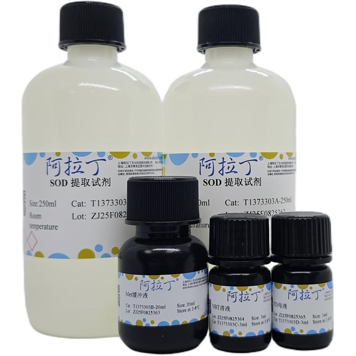
产品组分表:
| T1373303 | Component | 100 T | Storage | | T1373303A | SOD 提取试剂 | 2×250ml | RT | | T1373303B | Met缓冲液 | 20ml | 2-8 ℃. Store in the dark. | | T1373303C | NBT溶液 | 3ml | 2-8 ℃. Store in the dark. | | T1373303D | FD母液 | 3ml | 2-8 ℃. Store in the dark. |
|
自备材料:
1、生理盐水或PBS、去离子水
2、电子天平、剪刀、低温冰箱或制冰机、冰袋、匀浆器或研钵、离心机、离心管、小试管、40W荧光灯或日光灯、测光仪(照度计)、酶标仪、96孔板
操作步骤(仅供参考):
1、样品处理:
① 血浆或含红细胞的样品:从待测样品中分理出的血清或血浆不应有溶血,如果含有应去除红细胞后检测,如超过检测范围,用SOD提取试剂稀释后检测;血清去除红细胞的简易方法如下:用抗凝管收集血液,颠倒混匀,取至少500μl全血,4℃ 3000r/min离心5min,转移上清至另一新的1ml离心管中,适量生理盐水稀释后待测,亦可采用红细胞裂解液去除红细胞,如 ACK红细胞裂解液等。
② 组织样品:动物用含有20U/ml Heparin的生理盐水(0.9% NaCl containing 20U/ml Heparin)灌流清除血液后获取组织样品,按照每100mg组织加入500μl SOD提取试剂的比例,用玻璃匀浆器在4℃或冰浴匀浆,4℃ 4000r/min离心10min,取上清液(SOD粗提液)用于酶活性的测定。
③ 细胞样品:对于贴壁细胞,由于后续用于酶活性的测定,避免使用胰酶消化细胞,可以使用细胞刮或EDTA处理细胞并收集细胞,细胞用无菌的PBS或生理盐水洗涤1次,按照每106细胞加入300~500μl SOD提取试剂的比例,用玻璃匀浆器在4℃或冰浴匀浆,4℃ 10000r/min离心10min,取上清液(SOD粗提液)用于酶活性的测定。
④ 植物样品:准确称取植物材料(果肉或者去叶脉的叶片)0.4g,剪碎,置于4℃预冷的研钵或匀浆器中,加入预冷SOD提取试剂1ml,低温研磨至匀浆后转移至离心管,用3mlSOD提取试剂冲洗研钵或匀浆器并转入离心管,加提取试剂至总体积为4ml,4℃4000r/min 离心20min,上清液为酶提取液,上清液可用于SOD的检测。注意:如果SOD 酶活性较低,应相应减少提取试剂的总体积,以便提高SOD酶的浓度。
⑤ 上述样品准备完毕后可以用BCA法测定蛋白浓度,通常10~20μg蛋白的细胞或组织匀浆液样品其中的SOD平均活力约1个活力单位(不同细胞和组织的差异会比较大,该活力范围仅作为初步的参考);每种样品准备20~100μg蛋白量通常已经足够用于后续检测,根据蛋白浓度和预计的蛋白使用量,用该试剂盒提供的SOD提取试剂适当稀释样品,例如小鼠肝脏组织10%匀浆液(组织和匀浆液的重量比为10%)上清,通常需要稀释10~100倍,准备好的样品如果当天测定,可以冰浴保存;如果当天不能完成测定,可以-20℃冻存,但建议尽量当天完成测定。
2、(选做)准备SOD标准品:需自备SOD标准品,用本试剂盒提供的SOD提取试剂将SOD标准品稀释至如下系列浓度:200、100、50、20、10、5、2U/ml,各取20μl参考样品进行检测。注意:为避免稀释后SOD酶活性的下降,SOD标准品宜现稀释现使用,本试剂盒对于SOD的检测并不需要SOD作为标准品,但可使用SOD标准品作为阳性对照或作为对SOD活性定量的参考。
3、选取合适的光源:在光照培养箱或日光灯下,用光度仪测定光度值为3500~4000Lx的适合光照反应的位置,做出标记。
4、配制NBT工作液:将Met缓冲液与NBT溶液按23:3的比例混合即可。
5、配制FD工作液:将FD母液和去离子水按2:1~2:3的比例混合,466nm处吸光度为0.30~0.45即可使用,如果不在此范围,可通过增加或减少去离子水的加入量来调节。
6、SOD加样:参考下表使用96孔板设置空白对照孔、光照对照孔、测定孔,在低光强条件下按下表依次加入待测样品和其它各种溶液,加入FD工作液后充分混匀。注意:加入FD溶液后反应即会开始,在低光强条件下用排枪操作可减小各孔间因加入试剂的时间先后差异而导致的误差。
加入物(μl) | 空白对照孔 | 光照对照孔 | 测定孔 | SOD提取试剂 | 8 | 8 | | 待测样品(上清液) | | | 8 | NBT 工作液 | 208 | 208 | 208 | FD工作液 | 24 | 24 | 24 |
|
7、SOD测定:混匀,取空白对照孔置于暗处,其他各孔置于4000Lx日光下反应20min,各孔受光情况应一致,温度高时时间可缩短,低时延长;反应结束后,立即以不照光的空白对照孔调零,用酶标仪测定560nm处吸光度。
计算:
SOD活力单位的定义:以抑制NBT光化还原的50%为一个酶活性单位(U)。
液体中总SOD活力(U/ml)=(A光照-A测定)×V₁/(50%×A光照×VT)
组织、细胞匀浆液中总SOD活力(U/g)=(A光照-A测定)×V/(50%×A光照×VT×W)
血液中总SOD活力(U/gHb)=(A光照-A测定)×V×C/(50%×A光照×Hb)
式中:
A光照=光照对照孔的吸光度
A测定=测定孔的吸光度
V=样品液总体积(ml)
VT=测定时样品所用体积(ml)
W=样品鲜质量(g)
C=1ml/采血量(ml)
Hb=血红蛋白含量(gHb/ml)
注意事项:
1、上述低温试剂避免反复冻融,以免失效或效率下降。待测样品-70℃可保存1个月,需注意反复冻融会导致SOD部分失活。
2、植物中的多酚类物质会引起酶蛋白不可逆沉淀,使酶失去活性,因此在提取SOD酶时,必须添加多酚类物质的吸附剂,将多酚物质除去,避免酶蛋白变性失活。SOD提取试剂含有PVP等成分,可有效去除多酚类物质。SOD提取试剂不够用可以用磷酸钠缓冲液(0.05M,pH7.8)替代。
3、细胞或组织等样品制备时,不能采用含有Triton X-100等去垢剂的溶液,否则会干扰本试剂盒的检测。
4、抗氧化物会对本试剂盒的检测产生干扰,例如0.1mM ascorbic acid,5mM GSH都会使测定出来的吸光度显著升高。
5、对于植物样品,研磨处理应迅速,以免SOD酶活下降,尽量在冰浴条件下处理。
6、如果用分光光度计,比色杯光径应为1cm,加入的上清及试剂量应根据比色杯的最小要求体积而定。
7、所用离心管或96孔板应洁净透明,透光性好。
8、一般要求各孔受光情况一致,所有反应管应排列在与日光灯灯管平行的直线上。
9、反应温度控制在25℃,视酶活性高低适当调整反应时间;温度较高时,光照时间应缩短;温度较低时,光照时间相应延长。
10、如果无4000Lx日光,可200W在10~12cm处,照射20min;超净工作台5cm处光强约800~1000Lx,,照射30~40min;强太阳光下一般光强可达30000Lx,照射5~15min,一般不建议直接用太阳光。
11、为了您的安全和健康,请穿实验服并戴一次性手套操作。
12、试剂开封后请尽快使用,以防影响后续实验效果。
有效期:12个月有效。低温运输,按要求保存。
Superoxide Dismutase (SOD) is a metalloenzyme that catalyzes the dismutation of superoxide anion into hydrogen peroxide (H₂O₂) and oxygen (O₂). It is an essential antioxidant enzyme in organisms. Since superoxide radicals are unstable and have an extremely short lifespan, the activity of SOD is generally determined by indirect methods combined with various color reactions. Common chromogenic agents include Nitro Blue Tetrazolium (NBT), WST-1 and WST-8.
This product adopts the NBT-based photoreduction principle, also known as the NBT photoreduction method. The detection mechanism is as follows: riboflavin undergoes photoreduction in the presence of oxidizing substances. Under aerobic conditions, the reduced riboflavin is readily re-oxidized to generate superoxide anion radicals. These radicals reduce NBT into blue formazan, which has a strong absorbance at 560 nm. SOD scavenges superoxide anions and thus inhibits the formation of formazan.After the photoreduction reaction, the darker the blue color of the reaction solution, the lower the SOD activity; conversely, the higher the enzyme activity. The total SOD activity in samples can be calculated via colorimetric analysis using a microplate reader. This method quantifies SOD activity by measuring its inhibitory effect on the light-induced reduction of NBT. It is applicable to detect SOD activity in plants, tissues, cells, serum and other specimens. This kit is intended for research use only and is not for clinical diagnosis or other applications.
Product Components
| T1373303 | Component | 100 T | Storage | | T1373303A | SOD Extraction Reagent
| 2×250ml | RT | T1373303B
| Met buffer solution
| 20ml | 2-8 ℃. Store in the dark. | | T1373303C | NBT solution
| 3ml | 2-8 ℃. Store in the dark. | | T1373303D | FD Stock Solution
| 3ml | 2-8 ℃. Store in the dark. |
|
Materials to be Prepared by User
1. Normal saline or PBS, deionized water.
2. Electronic balance, scissors, low-temperature refrigerator or ice maker, ice packs, homogenizer or mortar, centrifuge, centrifuge tubes, test tubes, 40W fluorescent lamp, illuminance meter, microplate reader, 96-well plate.
Operating Procedures (For Reference Only)
1. Sample Preparation
① Plasma or erythrocyte-containing samples
The separated serum or plasma shall be free of hemolysis. If hemolysis occurs, remove erythrocytes before detection. If the sample value exceeds the detection range, dilute it with SOD extraction reagent prior to testing.
Simple method for removing erythrocytes from serum: Collect blood using an anticoagulant tube and mix by inverting. Take no less than 500 μL whole blood, centrifuge at 3000 rpm for 5 minutes at 4 °C. Transfer the supernatant to a new 1 mL centrifuge tube, dilute with an appropriate amount of normal saline for later detection. Erythrocyte lysis solution (e.g., ACK lysis buffer) can also be used to remove erythrocytes.
② Tissue samples
Perfuse animal tissues with 0.9% NaCl normal saline containing 20 U/mL Heparin to eliminate residual blood before sampling. Add 500 μL SOD extraction reagent per 100 mg tissue. Homogenize the sample with a glass homogenizer on ice or at 4 °C. Centrifuge the homogenate at 4000 rpm for 10 minutes at 4 °C, and collect the supernatant (crude SOD extract) for activity assay.
③ Cell samples
For adherent cells, avoid trypsin digestion to preserve enzyme activity. Harvest cells using a cell scraper or EDTA treatment. Rinse cells once with sterile PBS or normal saline. Add 300–500 μL SOD extraction reagent per 10⁶ cells, and homogenize with a glass homogenizer on ice or at 4 °C. Centrifuge at 10000 rpm for 10 minutes at 4 °C, then collect the supernatant (crude SOD extract) for activity assay.
④ Plant samples
Accurately weigh 0.4 g plant material (pulp or leaf blades with veins removed) and cut into pieces. Place the sample in a pre-chilled mortar or homogenizer at 4 °C, add 1 mL pre-cooled SOD extraction reagent, and grind thoroughly on ice. Transfer the homogenate to a centrifuge tube. Rinse the mortar or homogenizer with 3 mL SOD extraction reagent and combine the rinse solution into the same tube. Top up with extraction reagent to a total volume of 4 mL. Centrifuge at 4000 rpm for 20 minutes at 4 °C; the resulting supernatant is the enzyme extract for SOD detection.
Note: If the SOD activity is low, reduce the total volume of extraction reagent to increase enzyme concentration.
⑤ Post-treatment of prepared samples
Determine the protein concentration of samples via the BCA method. Generally, 10–20 μg protein from cell or tissue homogenate contains approximately 1 unit of SOD activity (values vary greatly across cell and tissue types, for reference only). A protein content of 20–100 μg per sample is sufficient for subsequent detection.
Dilute samples appropriately with the supplied SOD extraction reagent according to protein concentration and required dosage. For example, 10% mouse liver tissue homogenate (weight ratio of tissue to homogenate: 10%) usually requires a dilution ratio of 10–100 times. Store prepared samples on ice if assayed on the same day. For delayed detection, freeze samples at -20 °C. Assay samples on the day of preparation whenever possible.
2. (Optional) Preparation of SOD Standard
Prepare a series of SOD standard solutions at concentrations of 200, 100, 50, 20, 10, 5 and 2 U/mL using the supplied SOD extraction reagent. Pipette 20 μL of each standard solution for detection.
Note: Prepare and use the standard solutions immediately to prevent activity loss. SOD standard is not mandatory for this assay, but it can serve as a positive control or reference for quantitative analysis.
3. Selection of Light Source
Use an illuminance meter to identify positions with a light intensity of 3500–4000 Lux in a light incubator or under fluorescent lamps for the photoreaction, and mark the positions.
4. Preparation of NBT Working Solution
Mix Met buffer and NBT solution at a volume ratio of 23:3.
5. Preparation of FD Working Solution
Mix FD stock solution with deionized water at a volume ratio of 2:1 to 2:3. The solution is ready for use when its absorbance at 466 nm reaches 0.30–0.45. Adjust the volume of deionized water to bring the absorbance into the target range if needed.
6. Sample Loading for SOD Assay
Set up blank control wells, light control wells and test wells in a 96-well plate as specified in the attached table. Add test samples and other reagents sequentially under low light intensity, and mix thoroughly after adding FD working solution.
Note: The reaction starts immediately after FD working solution is added. Use a multi-channel pipette under low light to minimize errors caused by inconsistent reagent adding time across wells.
Additive (μL) | Blank control well | Light control well | Test well | SOD Extraction Reagent | 8 | 8 | - | Test sample (supernatant) | - | - | 8 | NBT working solution | 208 | 208 | 208 | FD working solution | 24 | 24 | 24 |
|
7. SOD Measurement
Mix the solutions thoroughly. Place the blank control wells in the dark, and incubate all other wells under a 4000 Lux fluorescent lamp for 20 minutes. Ensure uniform light exposure for all wells. Adjust the reaction duration: shorten it at high temperatures and extend it at low temperatures. After the reaction, immediately set the blank control wells (without light exposure) as the zero reference, then measure the absorbance at 560 nm using a microplate reader.
Calculation
Definition of SOD Activity Unit: One unit (U) of enzyme activity is defined as the amount of SOD causing 50% inhibition of the photochemical reduction of NBT.
Total SOD activity in liquid (U/mL)=(Alight control−Atest) ×V₁/ (50%×Alight control×VT)
Total SOD activity in tissue and cell homogenate (U/g)=(Alight control−Atest)×V₁×V/ (50%×Alight control×VT×W)
Parameter explanation
· A<sub>light control</sub>: Absorbance of light control well
· Atest: Absorbance of test well
· V₁: Total volume of reaction solution (mL)
· V: Total volume of sample solution (mL)
· VT: Volume of sample used in detection (mL)
· W: Fresh weight of sample (g)
· C: 1 mL/Volume of blood collected (mL)
Precautions
1. Do not repeatedly freeze and thaw low-temperature reagents, as this may cause reagent failure or reduced performance. Test samples can be stored at -70 °C for one month. Repeated freeze-thaw cycles will partially inactivate SOD.
2. Polyphenols in plant samples can trigger irreversible precipitation of enzyme proteins and lead to enzyme deactivation. Therefore, polyphenol adsorbents must be added during SOD extraction to remove such substances and prevent protein denaturation. The supplied SOD extraction reagent contains PVP and other components to effectively eliminate polyphenols. If the extraction reagent is insufficient, 0.05 M sodium phosphate buffer (pH 7.8) can be used as a substitute.
3. Avoid solutions containing detergents such as Triton X-100 during the preparation of cell and tissue samples, which will interfere with the assay results.
4. Antioxidants may interfere with the detection. For instance, 0.1 mM ascorbic acid and 5 mM GSH will significantly increase the measured absorbance values.
5. Grind plant samples quickly on ice to prevent the decline of SOD activity.
6. If a spectrophotometer is used, cuvettes with a 1 cm optical path length are required. The volumes of supernatant and reagents added shall comply with the minimum volume requirement of the cuvettes.
7. Use clean, transparent centrifuge tubes or 96-well plates with good light transmittance.
8. Ensure uniform light exposure for all wells. Arrange all reaction vessels in a line parallel to the fluorescent lamp tube.
9. Maintain the reaction temperature at 25 °C, and adjust the reaction time according to SOD activity. Shorten the illumination time at higher temperatures and extend it at lower temperatures.
10. If a 4000 Lux light source is unavailable, alternative light conditions are as follows: Use a 200 W lamp at a distance of 10–12 cm for 20 minutes; The light intensity is about 800–1000 Lux at 5 cm above a clean bench, with an illumination time of 30–40 minutes. Direct strong sunlight (approximately 30000 Lux) only requires 5–15 minutes of irradiation, which is not recommended.
11. Wear lab coats and disposable gloves during operation for personal safety and health.
12. Use opened reagents promptly to avoid affecting subsequent experiments.

 J2620429

 危险品化学品经营许可证(带存储)