谷胱甘肽过氧化物酶(Glutathione Peroxidase,GSH-Px)是一种含硒的水溶性四聚体蛋白酶,几乎在所有组织中都有分布,在一些病理状况下谷胱甘肽过氧化物酶的活力会发生明显的变化,该酶可以清除活细胞内过氧化物,在保护细胞免受自由基损伤过程中起着关键作用,细胞内的脂类容易和自由基发生反应,产生脂类过氧化物;谷胱甘肽过氧化物酶不仅具有消除自由基和衍生物的作用,还与过氧化氢酶(CAT)、磷脂氢过氧化物谷胱甘肽过氧化物酶(PH-GSH-Px)、谷胱甘肽S转移酶(GST)构成不同基质特异性的多水平的还原有机氢过氧化物系统,减少脂质过氧化物的形成,增强机体抗氧化损伤能力。
检测原理:GSH-Px可以促使过氧化氢(H₂O₂)与还原型谷胱甘肽(GSH)反应生成H₂O及氧化型谷胱甘肽(GSSG),谷胱甘肽过氧化物酶的活力可用其酶促反应的速度来表示,测定此酶促反应中还原型谷胱甘肽的消耗量,则可求出酶的活力。GSH-Px的活力以催化GSH的反应速度来表示,由于这两个底物在没有酶的条件下,也能进行氧化还原反应(称为非酶促反应),所以最后计算此酶活力时必须扣除非酶促反应引起的GSH减少的部分。而GSH含量的测定可以跟据GSH和5,5’-二硫代对硝基苯甲酸(DTNB)作用生成稳定黄色的5-硫代-2-硝基苯甲酸阴离子,在412nm处测其吸光度变化即可测出谷胱甘肽过氧化物酶的活力。
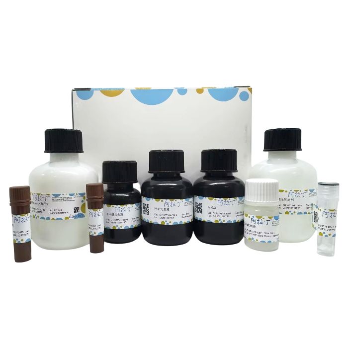
产品组分表
G1505754
| Component
| 50T
| Storage
|
G1505754A
| 样品匀浆液 | 50 mL | RT |
G1505754B
| GSH | 15.4 mg | 2-8℃ |
G1505754C
| GSH配制液 | 10 mL | RT
|
G1505754D
| 氧化剂 | 1 mL×2 | 2-8℃. Store in the dark.
|
G1505754E
| 酸性沉淀剂 | 100 mL
| RT
|
G1505754F
| GSH-Px Assay Buffer | 62.5 mL
| RT
|
G1505754G
| 苯甲酸显色液 | 15 mL
| -20℃. Store in the dark.
|
G1505754H
| ddH₂O | 50 mL
| RT |
自备仪器和试剂
1、生理盐水或PBS
2、离心管或EP管、分光光度计、比色皿、水浴锅或恒温箱、离心机
实验流程
1、样本处理
1.1 血清、血浆样本
从待测样本中分离出的血清或血浆不应有溶血,如果含有,应去除红细胞后检测,如超过检测范围,用生理盐水稀释后检测。血清去除红细胞的简易方法如下:用抗凝管收集血液,颠倒混匀,取至少500μl全血,4℃ 3000r/min离心5min,弃上清,用预冷的的10倍体积的样品匀浆液重悬红细胞沉淀,再次4℃ 3000r/min离心5min,弃上清,加入约4倍体积预冷的ddH₂O裂解红细胞沉淀,12000 r/min离心5min,取上清;亦可采用红细胞裂解液等去除红细胞,取上清。
1.2 组织样本
动物用含有20U/ml Heparin的生理盐水(0.9% NaCl containing 20U/ml Heparin)灌流清除血液后获取组织样品,按照每20mg组织加入200μl样品匀浆液的比例,用玻璃匀浆器在4℃或冰浴匀浆,4℃ 12000 r/min离心10min,取上清。
1.3 细胞样本
对于贴壁细胞,由于后续用于酶活性的测定,避免使用胰酶消化细胞,可用细胞刮或EDTA处理细胞收集细胞,细胞用PBS或生理盐水洗涤1次,按照每 10⁶ 细胞加入300~500μl匀浆液的比例用玻璃匀浆器在4℃或冰浴匀浆,4℃ 12000 r/min离心10min,取上清,用于酶活性的测定;亦可采用RIPA裂解液,参考相应说明裂解细胞样品,按照每 10⁶ 细胞加入100~200μl裂解液的比例进行裂解,取上清。
1.4 植物样本
称取0.2g新鲜样品或-80℃冻存的样品,放入预冷的研钵中,加入2ml预冷的磷酸缓冲液(0.05M,pH7.0),在冰浴上研磨或匀浆,转入离心管,4℃ 12000r/min 离心10~15min,取上清,用于酶活性的测定。
2、GSH工作液的配制
取0.5ml ddH₂O 加入15.4mg GSH中,充分溶解并混匀,即获得GSH储存液 (100mmol/L),立即分装后-20℃保存,取适量的GSH储存液 (100mmol/L),按GSH配制液:GSH储存液 (100mmol/L)=99:1的比例混合,即为GSH工作液 (1mmol/L),该溶液配制好以后可4℃保存1天。
3、氧化工作液的配制
准确取氧化剂0.1ml加入6.5ml ddH₂O,即为氧化储存液 (100×),4℃保存,临用前准确取氧化储存液 (100×) 0.1ml加入9.9ml ddH₂O,即为氧化工作液;4℃保存,1天有效。
4、GSH-Px酶促反应
参考下表,用离心管设置空白对照管、本底对照管、测定管,并按下表设置检测反应体系,依次加入试剂:
加入物质 (ml) | 空白对照管 | 本底对照管 | 测定管 |
GSH工作液 (1mmol/L)
| — | 0.2 | 0.2 |
待测样品 | — | — | 0.2 |
ddH₂O
| 0.2
| 0.2
| — |
混匀,置于37℃水浴5min。
混匀,置于37℃水浴5min。
3500g离心10min。
5、GSH-Px显色反应
参考下表,用96孔板或离心管设置空白对照管、本底对照管、测定管,并按下表设置检测反应体系,依次加入试剂:
加入物质 (ml)
| 空白对照管
| 本底对照管
| 测定管
|
取上清液
| —
| 1 | 1
|
空白对照 | 1
| —
| —
|
GSH-Px Assay Buffer | 1.25 | 1.25 | 1.25 |
苯甲酸显色液 | 0.25 | 0.25 | 0.25 |
6、GSH-Px测定
混匀,
置于室温孵育1min,以ddH₂O调零,用分光光度计或酶标仪检测412nm处吸光度(即为A空白、A本底、A测定);如果用分光光度计,比色杯光径应为1cm,加入的量应根据比色杯的最小量程而定;如果用酶标仪,96孔板每孔应加250μl;经测定,一般情况下A空白在0.003~0.05之间,A本底在0.1~0.3左右。
7、计算
谷胱甘肽过氧化物酶活力单位的定义:排除非酶促反应,在37℃,每1L血清, 1min内可以催化1μmol GSH氧化所需(减少)的酶量为一个GSH-Px活性单位。
GSH-Px (U/L)=(A本底-A测定)/(A本底-A空白)×200
参数说明:
A空白:空白对照的吸光度
A本底:本底对照的吸光度
A测定:待测样品的吸光度
200:1000(ml)/5(min)
注:
① [检测体系中谷胱甘肽过氧化物酶活力]的单位为U/L=mU/ml。
② [样品(如组织样本)中谷胱甘肽过氧化物酶活力]=[检测体系中谷胱甘肽过氧化物酶活力]×[稀释倍数]/[样品中的蛋白浓度]
[样品中(如组织样本)谷胱甘肽过氧化物酶活力]的单位为:U/mg或mU/mg蛋白
[样品中的蛋白浓度]的单位为:mg/ml。
③ 计算示例:样品的蛋白浓度经测定为0.5mg/ml,稀释2倍后进行测定。一般情况下,A空白在0.003~0.05之间,A本底在0.1~0.3左右。如果A本底=0.30,A测定=0.20,A空白=0.003;那么:
样本GSP-Px活力=(0.30-0.20)/(0.30-0.003)×200=67.34U/L
样本GSP-Px活力=67.34U/L×2/(0.5mg/ml)=269.36mU/mg (蛋白)
参考区间:成年人血清GSP-Px:115~140 U/L
注意事项
1、上述低温试剂避免反复冻融,以免失效或效率下降。
2、本法中所有氧化剂或还原剂都会干扰本试剂盒的测定,如果在样品中的还原剂无法避免,例如DTT、巯基乙醇等,则这些还原剂的总浓度至少低于0.1mM;0.15mM的DTT可以抑制40%的酶活力。
3、常用的Triton X-100、Tween 20等去垢剂都含有较高水平的过氧化物,会影响本试剂盒的测定,如果必须使用这些去垢剂,最好使用纯度较高并注明含较低浓度过氧化物的去垢剂。
4、样品取出后最好立即测定,也可以-80℃冻存待以后测定。
5、一定要严格控制反应时的温度,否则会引起较多误差。
6、试剂开封后请尽快使用,以防影响后续实验效果。
7、为了您的安全和健康,请穿实验服并戴一次性手套操作。
Glutathione Peroxidase (GSH-Px) is a selenium-containing, water-soluble tetrameric proteolytic enzyme distributed in almost all tissues. Its activity can change significantly under certain pathological conditions. This enzyme scavenges peroxides within living cells and plays a key role in protecting cells from free radical damage. Intracellular lipids are prone to react with free radicals, producing lipid peroxides. GSH-Px not only eliminates free radicals and their derivatives but also, together with Catalase (CAT), Phospholipid Hydroperoxide Glutathione Peroxidase (PH-GSH-Px), and Glutathione S-Transferase (GST), forms a multi-level system for reducing organic hydroperoxides with different substrate specificities. This system reduces the formation of lipid peroxides and enhances the body's capacity to resist oxidative damage.
Assay Principle
GSH-Px catalyzes the reaction between hydrogen peroxide (H₂O₂) and reduced glutathione (GSH) to produce H₂O and oxidized glutathione (GSSG). The activity of GSH-Px can be represented by the rate of this enzymatic reaction. By measuring the consumption of reduced glutathione in this reaction, the enzyme activity can be determined. The activity of GSH-Px is expressed as the rate of GSH reaction. Since these two substrates can also undergo redox reactions without the enzyme (non-enzymatic reaction), the final calculation of enzyme activity must subtract the portion of GSH reduction caused by the non-enzymatic reaction. The GSH content is measured based on the reaction between GSH and 5,5'-Dithiobis(2-nitrobenzoic acid) (DTNB), which produces a stable yellow 5-thio-2-nitrobenzoic acid anion. The change in absorbance at 412 nm is used to determine Glutathione Peroxidase activity.
G1505754
| Component
| 50T
| Storage
|
G1505754A
| Sample Homogenization Buffer
| 50 mL | RT |
G1505754B
| GSH | 15.4 mg | 2-8℃ |
G1505754C
| GSH Preparation Solution
| 10 mL | RT
|
G1505754D
| Oxidizing Agent
| 1 mL×2 | 2-8℃. Store in the dark.
|
G1505754E
| Acidic Precipitation Agent
| 100 mL
| RT
|
G1505754F
| GSH-Px Assay Buffer | 62.5 mL
| RT
|
| G1505754G | Benzoic Acid Chromogenic Solution
| 15 mL
| -20℃. Store in the dark. |
Required Materials Not Provided
1. Physiological saline or PBS
2. Centrifuge tubes or EP tubes, Spectrophotometer, Cuvettes, Water bath or incubator, Centrifuge
Experimental Procedure
1. Sample Preparation
1.1 Serum/Plasma Samples
Serum or plasma separated from the sample should not be hemolyzed. If present, remove red blood cells before assay. If exceeding the detection range, dilute with physiological saline before assay.
Simple method for RBC removal from serum: Collect blood in an anticoagulant tube, mix by inverting. Take at least 500 µL whole blood, centrifuge at 3000 rpm/min for 5 min at 4°C, discard supernatant. Resuspend the RBC pellet in 10 volumes of pre-chilled Sample Homogenization Buffer, centrifuge again (3000 rpm/min, 5 min, 4°C), discard supernatant. Add approx. 4 volumes of pre-chilled ddH₂O to lyse the RBC pellet, centrifuge at 12,000 rpm/min for 5 min, collect supernatant. Alternatively, use products like ACK Lysis Buffer to remove RBCs, then collect supernatant.
1.2 Tissue Samples
Perfuse animals with physiological saline containing 20 U/ml Heparin to remove blood before collecting tissue samples.
Homogenize tissue on ice or at 4°C using a glass homogenizer with 200 µL Sample Homogenization Buffer per 20 mg tissue.
Centrifuge at 12,000 rpm/min for 10 min at 4°C. Collect the supernatant.
1.3 Cell Samples
For adherent cells: Avoid trypsin digestion as it may affect subsequent enzyme activity assays. Use a cell scraper or EDTA to harvest cells. Wash cells once with PBS or physiological saline.
Homogenize cells on ice or at 4°C using a glass homogenizer with 300-500 µL Homogenization Buffer per 10⁶ cells.
Centrifuge at 12,000 rpm/min for 10 min at 4°C. Collect the supernatant for enzyme assay.
Alternatively: Lyse cells using RIPA Lysis Buffer according to the manufacturer's instructions, using 100-200 µL lysis buffer per 10⁶ cells. Collect the supernatant.
1.4 Plant Samples
Weigh 0.2 g of fresh or -80°C frozen sample, place in a pre-chilled mortar.
Add 2 mL of pre-chilled Phosphate Buffer (0.05 M, pH 7.0).
Grind or homogenize on ice.
Transfer to a centrifuge tube, centrifuge at 12,000 rpm/min for 10-15 min at 4°C.
Collect the supernatant for enzyme assay.
2. Preparation of GSH Working Solution
Add 0.5 mL ddH₂O to the 15.4 mg GSH vial. Dissolve and mix thoroughly to obtain the GSH Stock Solution (100 mmol/L).
Aliquot immediately and store at -20°C.
For the GSH Working Solution (1 mmol/L), mix GSH Preparation Solution and GSH Stock Solution (100 mmol/L) at a 99:1 ratio.
This working solution can be stored at 4°C for 1 day after preparation.
3. Preparation of Oxidizing Working Solution
Accurately pipette 0.1 mL of the Oxidizing Agent into 6.5 mL ddH₂O to prepare the Oxidizing Stock Solution (100X). Store at 4°C.
Before use, accurately pipette 0.1 mL of the Oxidizing Stock Solution (100X) into 9.9 mL ddH₂O to prepare the Oxidizing Working Solution.
Store at 4°C, stable for 1 day.
4. GSH-Px Enzymatic Reaction
Set up Blank Control, Background Control, and Test tubes in centrifuge tubes according to the table below.
Add reagents in the specified order:
Reagent (mL) | Blank Control Tube | Background Control Tube
| Test Tube |
GSH Working Solution (1 mmol/L)
| — | 0.2 | 0.2 |
Sample | — | — | 0.2 |
ddH₂O
| 0.2
| 0.2
| — |
Mix well and incubate at 37°C for 5 min.
Oxidizing Working Solution (pre-warmed 37°C) | —
| 0.1
| 0.1 |
Mix well and incubate at 37°C for 5 min.
Acidic Precipitation Agent | 0.8 | 2.0 | 2.0 |
Centrifuge at 3500 g for 10 min.
Supernatant Collected | — | 1.0 | 1.0 |
5. GSH-Px Chromogenic Reaction
Set up Blank Control, Background Control, and Test wells/tubes in a 96-well plate or centrifuge tubes according to the table below.
Add reagents in the specified order:
Reagent (mL)
| Blank Control Tube
| Background Control Tube
| Test Tube
|
Supernatant from Step 4
| —
| 1.0 | 1.0
|
Blank Control from Step 4 | 1.0
| —
| —
|
GSH-Px Assay Buffer | 1.25 | 1.25 | 1.25 |
Benzoic Acid Chromogenic Solution | 0.25 | 0.25 | 0.25 |
6. GSH-Px Measurement
Mix well and incubate at room temperature for 1 minute.
Zero the instrument with ddH₂O.
Measure the absorbance at 412 nm using a spectrophotometer or microplate reader (recorded as ABlank, ABackground, ATest).
Note for spectrophotometers: Use a 1 cm light path cuvette; adjust volumes based on the cuvette's minimum requirement.
Note for microplate readers: Add 250 µL per well if using a 96-well plate.
Typically, ABlank ranges from 0.003 to 0.05, and ABackground ranges from 0.1 to 0.3.
7. Calculation
Definition of GSH-Px Activity Unit: Excluding non-enzymatic reactions, one unit of GSH-Px activity is defined as the amount of enzyme required to catalyze the oxidation (consumption) of 1 μmol of GSH per minute per liter of serum at 37°C.
GSH-Px (U/L) = (ABackground - ATest) / (ABackground - ABlank) × 200
Parameter Definitions:
ABlank: Absorbance of the Blank Control
ABackground: Absorbance of the Background Control
ATest: Absorbance of the Test sample
200: Derived from 1000 (mL) / 5 (min) – relates unit definition to assay conditions.
Notes:
① The unit for [Glutathione Peroxidase activity in the detection system] is U/L, which is equivalent to mU/mL.
② [Glutathione Peroxidase activity in the sample (e.g., tissue)] = [GSH-Px activity in detection system] × [Dilution Factor] / [Sample Protein Concentration]
* Unit for [GSH-Px activity in sample]: U/mg or mU/mg protein
* Unit for [Sample Protein Concentration]: mg/mL
③ Calculation Example:
* Sample protein concentration: 0.5 mg/mL, assayed after a 2-fold dilution.
* Typical values: ABlank ~0.003-0.05, ABackground ~0.1-0.3.
* If ABackground = 0.30, ATest = 0.20, ABlank = 0.003:
* Sample GSH-Px activity = (0.30 - 0.20) / (0.30 - 0.003) × 200 = 67.34 U/L
* Sample GSH-Px activity = 67.34 U/L × 2 / (0.5 mg/mL) = 269.36 mU/mg protein
Reference Interval: Adult Serum GSH-Px: 115 ~ 140 U/L
Precautions
Avoid repeated freeze-thaw cycles for the low-temperature reagents to prevent inactivation or reduced efficiency.
All oxidizing or reducing agents can interfere with this assay. If unavoidable in the sample (e.g., DTT, β-mercaptoethanol), keep the total concentration of such reductants below 0.1 mM. Note: 0.15 mM DTT can inhibit 40% of enzyme activity.
Common detergents like Triton X-100 and Tween 20 often contain high levels of peroxides, which can interfere. If necessary, use high-purity grades specified to have low peroxide content.
Assay samples immediately after preparation, or store at -80°C for later analysis.
Strictly control the reaction temperature to minimize errors.
Use reagents promptly after opening to avoid affecting subsequent experimental results.
For your safety and health, wear lab coats and disposable gloves during operation.

 H2614318


 危险品化学品经营许可证(带存储)