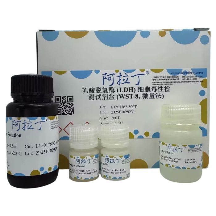
乳酸脱氢酶 (LDH) 细胞毒性检测试剂盒 (WST-8, 微量法) 是一种基于WST-8的显色反应,通过比色法检测细胞毒性时释放的乳酸脱氢酶(Lactate dehydrogenase, LDH)活性或检测其它样品中的LDH活性的试剂盒。
检测原理:LDH是绝大部分哺乳动物活细胞中都存在的酶,受损细胞所释放出的LDH催化乳酸(Lactate)生成丙酮酸(Pyruvate),并伴随着NAD⁺到NADH的转化。NADH通过电子载体可将水溶性四唑盐WST-8还原成水溶性的橙色甲臜产物(Formazan),在490nm波长下产生吸收峰,甲臜产物的吸光度与LDH的量呈正相关,利用此原理可以测定死细胞和受损细胞的数量。该酶联反应原理的示意图如下。

本试剂盒所采用的WST-8法跟经典的INT法相比,检测反应迅速、灵敏度更高,吸光度变化更大,检测结果更准确。可以用于常规的LDH活性的检测,尤其适用于以LDH释放为指标的细胞毒性/损伤检测。同时,基于细胞裂解后LDH活性的检测,本试剂盒也可以用于检测细胞增殖。本试剂盒搭配CCK-8(货号C266180/S707171)检测,可作为细胞毒性双指标认证,更适合Car-T、ADCC、CDC等效靶杀伤实验。
本试剂盒中试剂不会和活细胞反应,而且对细胞无损害,可以直接添加到含有细胞的培养基中检测 (一步法),也可以取细胞上清检测(低损伤法,分离的细胞可以做其他实验)。
L1501762
| Component
| 100T | 500T | Storage
|
L1501762A
| Lysis Buffer
| 1.1 mL | 5.5 mL
| -20℃. Store in the dark.
|
L1501762B
| Catalyst Solution
| 1.1 mL
| 5.5 mL
| -20℃. Store in the dark.
|
L1501762C
| Dye Solution
| 9.9 mL
| 49.5 mL | -20℃. Store in the dark.
|
L1501762D
| Stop Solution
| 5.5 mL
| 27.5 mL | -20℃. Store in the dark. |
产品优势
双重选择—有两种检测方法可供选择(一步法、低损伤法);
灵敏度高—反应迅速,吸光度变化大,检测结果准确;
操作便捷—使用一步法不需要离心;
双重验证—与CCK-8联用可更全面评估细胞活力和细胞损伤。
实验流程
1、实验前准备
1.1 分组
| 样品
| 样品Blank
| 高对照
| 高对照Blank
| 低对照
| 背景Blank
|
培养基
|
| √
| √
| √
| √
| √
|
细胞悬液
| √ |
| √
|
| √
|
|
药物
| √
| √
|
|
|
|
|
Lysis Buffer
|
|
| √
| √
|
|
|
注:培养基中动物血清中的LDH会增加背景吸收,建议设置一个Blank组对照来校正。
1.2 方法的选择
一步法:无需离心,更便捷,适用于不需要收集活细胞进行其它实验的LDH检测。
低损伤法:适用于需要收集活细胞进行其它实验的LDH检测。
1.3 96孔板的准备
一步法:贴壁细胞和悬浮细胞都使用平底96孔板。
低损伤法:贴壁细胞使用平底96孔板,悬浮细胞使用圆底或V底96孔板。
1.4 配制LDH检测工作液
按照1:9的体积比例将Catalyst Solution和Dye Solution均匀混合即配制成LDH检测工作液。按照每孔100μL的体积,根据待测定的样品数(含对照),参考下表在临检测前新鲜配制适量的LDH检测工作液。
| 1孔
| 10孔
| 20孔
| 50孔
|
Catalyst Solution
| 10μL
| 0.1mL
| 0.2mL
| 0.5mL
|
Dye Solution
| 90μL
| 0.9mL
| 1.8mL
| 4.5mL |
注:LDH检测工作液必须现配现用,短期可于-20℃或2-8℃保存2-3天。配制和使用过程中均要注 意适当避光。
2、制作LDH标准曲线(可选)
如果需要进行LDH的绝对定量,需自备不同浓度的LDH标准品(如0、65、125、250、500、1000 mU/mL)。如有必要,在后续实验中可以根据样品的LDH活性,对标准曲线的浓度范围进行适当调整。
3、检测方法1:一步法
3.1 预实验—细胞数最佳化(可选)
因为每种细胞的LDH量不同,推荐通过预实验确定高对照和低对照的吸光度差异最大的细胞数。
(1) 制备细胞悬液,细胞计数,调整细胞密度至5×10⁵ cells/mL。
(2) 向96孔板中每孔加入100μL培养基。
(3) 如图1所示向2列中加入100μL细胞悬液,按1/2比例向右依次和培养基混合等比稀释成一系列细胞浓度梯度(推荐使用多通道移液器进行操作),并准备高对照Blank和背景Blank,每组3个复孔。

图1. 预实验96孔板分组设置示意图
(4) 在细胞培养箱中培养一定时间,此时间设定应与之后的细胞毒性时间一致。
(5) 在高对照孔中加入10μL Lysis Buffer,在低对照孔和背景Blank孔中加入10μL培养基。
(6) 在细胞培养箱中培养30min。
(7) 在每孔中加入100μL检测工作液,采用包裹铝箔等方法避光,在室温反应,吸光度与反应时间成正比,建议5-30min内检测。
注:因为不同细胞差异较大,建议首次实验时每隔五分钟测定一次吸光度,以确定最佳反应时间。
(8) 在每孔中加入50μL Stop Solution后,立刻用酶标仪测定490nm的吸光度。
(9) 以吸光度为X轴,细胞密度为Y轴绘制线性曲线,根据以下要求选择最佳的细胞浓度:
A:高对照和低对照的吸光度之差>0.2;
B:在线性曲线上的该细胞浓度的吸光度<2.0。
3.2 细胞毒性检测
(1) 将最适细胞数的待检细胞加入到96孔板中,每孔50μL。
(2) 贴壁细胞培养过夜,换液后继续进行步骤3;悬浮细胞可以在适当静置后继续进行步骤3。
(3) 加入50μL含有药物的培养基(如表1)。
(4) 在细胞培养箱中培养合适的时间。
(5) 在高对照孔中加入10μL Lysis Buffer后,在样品、样品Blank、低对照孔和背景Blank孔中加入10 μL培养基,在细胞培养箱内培养30min。
(6) 在每个孔中加入100μL检测工作液后,在室温的条件下避光孵育。培养时间设定与预实验一致, 若没有做预实验,可以在5-30min每隔5min观察颜色,当各组橙黄色差异明显时即可。
(7) 在每孔中加入50μL Stop Solution后,立刻用酶标仪测定490nm的吸光度。
表1. 细胞毒性试验各分组每孔溶液量(一步法)
| 样品
| 样品Blank
| 高对照
| 高对照Blank
| 低对照
| 背景Blank |
培养基
| 10μL
| 60μL
| 50μL
| 100μL
| 60μL
| 110μL |
细胞悬液
| 50μL
|
| 50μL
|
| 50μL |
|
药物
| 50μL
| 50μL
|
|
|
|
|
Lysis Buffer
|
|
| 10μL
| 10μL |
|
|
4、检测方法2:低损伤法
4.1 预实验—细胞数最佳化(可选)
因为每种细胞的LDH量不同,推荐通过预实验确定高对照和低对照的吸光度差异最大的细胞数。
(1) 制备细胞悬液,细胞计数,调整细胞密度至5×10⁵ cells/mL。
(2) 向96孔板中每孔加入100μL培养基。
(3) 如图1所示向2列中加入100μL细胞悬液,按1/2比例向右依次和培养基混合等比稀释成一系列细胞浓度梯度(推荐使用多通道移液器进行操作),并准备高对照Blank和背景Blank(只有培养基),每组3个复孔。
(4) 在每孔中加入100μL培养基。
(5) 在细胞培养箱中培养一定时间,此时间设定应与之后的细胞毒性时间一致。
(6) 在高对照孔中加入20μL Lysis Buffer,在低对照孔和背景Blank孔中加入20μL培养基。
(7) 在细胞培养箱中培养30min。
(8) 96孔板250×g离心2min,使悬浮细胞沉淀。
(9) 从每个孔中吸取100μL上清液至新的96孔板中。
注:请小心吸取上清液,避免吸出细胞。
(10) 在每孔中加入100μL检测工作液,采用包裹铝箔等方法避光,在室温反应,吸光度与反应时间成正比,建议5-30min内检测。
注:因为不同细胞差异较大,建议首次实验时每隔五分钟测定一次吸光度,以确定最佳反应时间。
(11) 在每孔中加入50μL Stop Solution后,立刻用酶标仪测定490nm的吸光度。
(12) 以吸光度为X轴,细胞密度为Y轴绘制线性曲线,根据以下要求选择最佳的细胞浓度:
A:高对照和低对照的吸光度之差>0.2;
B:在线性曲线上的该细胞浓度的吸光度<2.0。
4.2 细胞毒性检测
(1) 将最适细胞数的待检细胞加入到96孔板中,每孔100μL。
(2) 贴壁细胞培养过夜,换液后继续进行步骤3;悬浮细胞可以在适当静置后继续进行步骤3。
(3) 加入100μL含有药物的培养基(如表2)。
(4) 在细胞培养箱中培养合适的时间。
(5) 在高对照孔中加入20μL Lysis Buffer后,在样品、样品Blank、低对照孔和背景Blank孔中加入20μL培养基,在细胞培养箱内培养30min。
(6) 96孔板250×g离心2min,使悬浮细胞沉淀。
(7) 从每个孔中吸取100μL上清液至新的96孔板中。
注:请小心吸取上清液,避免吸出细胞。
(8) 在每个孔中加入100μL检测工作液后,在室温的条件下避光孵育。培养时间设定与预实验一致,若没有做预实验,可以在5-30min每隔5min观察颜色,当各组橙黄色差异明显时即可。
(9) 在每孔中加入50μL Stop Solution后,立刻用酶标仪测定490nm的吸光度。
表2. 细胞毒性试验各分组每孔溶液量(低损伤法)
| 样品
| 样品Blank
| 高对照
| 高对照Blank
| 低对照
| 背景Blank
|
培养基
| 20μL
| 120μL
| 100μL
| 200μL
| 120μL
| 220μL
|
细胞悬液
| 100μL
|
| 100μL
|
| 100μL
|
|
药物
| 100μL
| 100μL
|
|
|
|
|
Lysis Buffer
|
|
| 20μL
| 20μL
|
|
|
5、数据整理与计算公式
5.1 计算“样品孔-样品Blank孔”、“高对照孔-高对照Blank孔”、“低对照孔-背景Blank孔”的吸光度,算出n=3的平均值。
5.2 细胞损伤率=[(A-C) / (B-C)] ×100%
A:样品的平均吸光度“样品孔-样品Blank孔”
B:高对照的平均吸光度“高对照孔-高对照Blank孔”
C:低对照的平均吸光度“低对照孔-背景Blank孔”
保存条件
-20℃避光保存,自生产之日起12个月有效。建议将试剂分装保存,避免反复冻融导致产品失效,干扰实验测定。试剂盒解冻后可以短期于2-8℃保存,3天内有效。
注意事项
1、动物血清中的LDH活性较高时,可以通过降低血清浓度来减小其中的LDH造成的背景吸收,一般有两种解决方法:
① 将血清浓度降低到5%会显著减小背景而不影响细胞活力。不推荐用1%BSA来代替血清检测细胞介导的细胞毒性。
② 使用灭活血清,这样血清中的LDH背景会大幅度下降。
2、待检样品准备好后建议当天完成测定,如果暂时无法测定,则请置于2-8℃,可放置2-3天。不建议置于-20℃,会使样品中部分LDH失活。
3、细胞过度生长、密度过高、状态不佳、离心速度过大、培养箱内外温差过大,都会造成细胞释放LDH 增加。不同种类、不同状态的细胞LDH的含量也存在一定差异。
4、未加终止液时,随着时间延长,吸光度会逐渐增大。加入终止液后,显色可以避光稳定保存48小时。
5、如果酶标仪没有490nm滤光片,也可以使用450nm滤光片,吸光度会高于490nm。
6、CCK-8 以细胞活性为指标,本试剂盒以游离的LDH为指标,两种指标检测存在差异性,二者联用,能更全面的表征细胞毒性。
7、如果待测体系中存在氧化还原物质则可能会干扰检测结果,还原性物质会使吸光度增加,氧化性物质会使吸光度减小,因此应需设法去除这些物质的影响。
8、当在培养箱内培养时,培养板最外一圈的孔最容易干燥挥发,由于体积不准确而增加误差。一般情况下,最外一圈的孔只加培养基,不作为测定孔用。
9、为了您的安全和健康,操作时请穿着实验服并佩戴一次性手套。
This kit is a WST-8-based colorimetric assay designed to measure Lactate Dehydrogenase (LDH) activity released during cytotoxicity or present in other samples via a colorimetric method.
Detection Principle: LDH is an enzyme present in almost all living mammalian cells. LDH released from damaged cells catalyzes the conversion of lactate to pyruvate, accompanied by the conversion of NAD⁺ to NADH. NADH, via an electron carrier, reduces the water-soluble tetrazolium salt WST-8 to a water-soluble orange formazan product. This formazan product has an absorption peak at 490 nm. The absorbance of the formazan product is directly proportional to the amount of LDH. This principle is used to determine the number of dead and damaged cells. A schematic diagram of this coupled enzymatic reaction is provided.

Compared to the classic INT method, the WST-8 method used in this kit offers faster reaction kinetics, higher sensitivity, greater absorbance changes, and more accurate results. It can be used for routine LDH activity detection and is particularly suitable for cytotoxicity/damage assays where LDH release is the indicator. Furthermore, based on LDH activity detection after cell lysis, this kit can also be used to assess cell proliferation. When used in combination with the CCK-8 Assay Kit (Cat. No. C266180/S707171), it provides dual-parameter validation of cytotoxicity, making it especially suitable for target cell killing experiments like Car-T, ADCC, and CDC.
The reagents in this kit do not react with viable cells and are non-toxic, allowing for direct addition to the cell culture medium for detection (One-Step Method) or for the assay of cell culture supernatant (Low-Damage Method, allowing isolated cells to be used for other experiments).
L1501762
| Component
| 100T | 500T | Storage
|
L1501762A
| Lysis Buffer
| 1.1 mL | 5.5 mL
| -20℃. Store in the dark.
|
L1501762B
| Catalyst Solution
| 1.1 mL
| 5.5 mL
| -20℃. Store in the dark.
|
L1501762C
| Dye Solution
| 9.9 mL
| 49.5 mL | -20℃. Store in the dark.
|
L1501762D
| Stop Solution
| 5.5 mL
| 27.5 mL | -20℃. Store in the dark. |
Product Advantages:
Dual Options: Two detection methods available (One-Step Method, Low-Damage Method).
High Sensitivity: Fast reaction, large absorbance change, accurate results.
Convenient Operation: One-Step Method requires no centrifugation.
Dual Verification: Combined use with CCK-8 allows more comprehensive assessment of cell viability and damage.
Experimental Procedure
1. Pre-experiment Preparation
1.1 Group Setup
|
Group
| Sample
| Sample Blank
| High Control
| High Control Blank
| Low Control
| Background Blank |
Medium
|
| √
| √
| √
| √
| √
|
Cell Suspension
| √ |
| √
|
| √
|
|
Drug/Compound
| √
| √
|
|
|
|
|
Lysis Buffer
|
|
| √
| √
|
|
|
Note: LDH activity in animal serum can increase background absorbance. It is recommended to set up a Blank control group for correction.
1.2 Method Selection
One-Step Method: No centrifugation required, more convenient. Suitable for LDH detection where viable cells do not need to be collected for other experiments.
Low-Damage Method: Suitable for LDH detection where viable cells need to be collected for other experiments.
1.3 96-well Plate Preparation
One-Step Method: Use flat-bottom 96-well plates for both adherent and suspension cells.
Low-Damage Method: Use flat-bottom plates for adherent cells; use round-bottom or V-bottom plates for suspension cells.
1.4 Preparation of LDH Detection Working Solution
Prepare the LDH Detection Working Solution immediately before use by uniformly mixing the Catalyst Solution and Dye Solution in a 1:9 volume ratio. Prepare a sufficient volume based on the number of samples (including controls), using 100 μL per well. Refer to the table below for guidance.
Number of Wells
| 1
| 10
| 20
| 50
|
Catalyst Solution
| 10 μL
| 0.1 mL
| 0.2 mL
| 0.5 mL
|
Dye Solution
| 90 μL
| 0.9 mL
| 1.8 mL
| 4.5 mL |
Note: The LDH Detection Working Solution must be prepared fresh. It can be stored short-term at -20°C or 2-8°C for 2-3 days. Protect from light during preparation and use.
2. LDH Standard Curve (Optional)
If absolute quantification of LDH is required, prepare LDH standards at different concentrations (e.g., 0, 65, 125, 250, 500, 1000 mU/mL). Adjust the concentration range in subsequent experiments based on sample LDH activity if necessary.
3. Detection Method 1: One-Step Method
3.1 Pre-experiment - Cell Number Optimization (Optional)
As LDH content varies by cell type, a pre-experiment is recommended to determine the cell number yielding the maximum absorbance difference between High and Low controls.
(1) Prepare cell suspension, count cells, and adjust density to 5×10⁵ cells/mL.
(2) Add 100 μL medium to each well of a 96-well plate.
(3) Add 100 μL cell suspension to two columns. Perform serial 1:2 dilutions across the plate with medium to create a cell concentration gradient. Include High Control Blank and Background Blank groups, with 3 replicates per group.
(4) Incubate in a cell culture incubator for a duration matching the intended cytotoxicity assay time.
(5) Add 10 μL Lysis Buffer to High Control wells. Add 10 μL medium to Low Control and Background Blank wells.
(6) Incubate in the cell culture incubator for 30 min.
(7) Add 100 μL Detection Working Solution to each well. Protect from light (e.g., with aluminum foil) and incubate at room temperature. Absorbance is proportional to reaction time; measure within 5-30 min.
Note: Due to cell type variations, measure absorbance every 5 minutes during the first experiment to determine the optimal reaction time.
(8) Add 50 μL Stop Solution to each well, then immediately measure absorbance at 490 nm.
(9) Plot a graph with Absorbance on the X-axis and Cell Density on the Y-axis. Select the optimal cell concentration based on:
A: Difference between High and Low control absorbance > 0.2.
B: Absorbance at that cell concentration on the linear curve < 2.0.
3.2 Cytotoxicity Assay
(1) Seed cells at the optimal density into a 96-well plate, 50 μL per well.
(2) Culture adherent cells overnight, then change medium before step 3. Suspension cells can proceed to step 3 after brief settling.
(3) Add 50 μL of drug-containing medium to appropriate wells (see Table 1).
(4) Incubate in a cell culture incubator for the desired duration.
(5) Add 10 μL Lysis Buffer to High Control wells. Add 10 μL medium to Sample, Sample Blank, Low Control, and Background Blank wells. Incubate in the cell culture incubator for 30 min.
(6) Add 100 μL Detection Working Solution to each well. Incubate at room temperature protected from light. Use the incubation time determined in the pre-experiment. If no pre-experiment was done, monitor color every 5-30 min; stop when distinct orange-yellow differences are observed between groups.
(7) Add 50 μL Stop Solution to each well, then immediately measure absorbance at 490 nm.
Table 1. Volume per Well for Cytotoxicity Test (One-Step Method)
Reagent
| Sample (μL) | Sample Blank (μL)
| High Control (μL)
| High Control Blank (μL)
| Low Control (μL)
| Background Blank (μL) |
Medium
| 10
| 60
| 50
| 100
| 60
| 110 |
Cell Suspension
| 50
|
| 50
|
| 50 |
|
| Drug/Compound | 50
| 50
|
|
|
|
|
Lysis Buffer
|
|
| 10
| 10 | |
4. Detection Method 2: Low-Damage Method
4.1 Pre-experiment - Cell Number Optimization (Optional)
(Follow steps 3.1 (1)-(4) for cell preparation, dilution series, and blanks)
(5) Add 100 μL medium to each well.
(6) Incubate in a cell culture incubator for a duration matching the intended cytotoxicity assay time.
(7) Add 20 μL Lysis Buffer to High Control wells. Add 20 μL medium to Low Control and Background Blank wells.
(8) Incubate in the cell culture incubator for 30 min.
(9) Centrifuge the plate at 250 × g for 2 min to pellet suspension cells.
(10) Carefully transfer 100 μL of supernatant from each well to a new 96-well plate.
Note: Pipette carefully to avoid disturbing the cell pellet.
(11) Add 100 μL Detection Working Solution to each well of the new plate. Protect from light and incubate at room temperature. Measure within 5-30 min.
Note: Determine optimal reaction time as in 3.1(7).
(12) Add 50 μL Stop Solution to each well, then immediately measure absorbance at 490 nm.
(13) Plot graph and select optimal cell concentration as in 3.1(9).
4.2 Cytotoxicity Assay
(1) Seed cells at the optimal density into a 96-well plate, 100 μL per well.
(2) Culture adherent cells overnight, then change medium before step 3. Suspension cells can proceed to step 3 after brief settling.
(3) Add 100 μL of drug-containing medium to appropriate wells (see Table 2).
(4) Incubate in a cell culture incubator for the desired duration.
(5) Add 20 μL Lysis Buffer to High Control wells. Add 20 μL medium to Sample, Sample Blank, Low Control, and Background Blank wells. Incubate in the cell culture incubator for 30 min.
(6) Centrifuge the plate at 250 × g for 2 min to pellet suspension cells.
(7) Carefully transfer 100 μL of supernatant from each well to a new 96-well plate.
Note: Pipette carefully to avoid disturbing the cell pellet.
(8) Add 100 μL Detection Working Solution to each well of the new plate. Incubate at room temperature protected from light. Use the pre-determined time or monitor color as before.
(9) Add 50 μL Stop Solution to each well, then immediately measure absorbance at 490 nm.
Table 2. Volume per Well for Cytotoxicity Test (Low-Damage Method)
Reagent
| Sample (μL)
| Sample Blank (μL)
| High Control (μL)
| High Control Blank (μL)
| Low Control (μL)
| Background Blank (μL)
|
Medium
| 20
| 120
| 100
| 200
| 120
| 220
|
Cell Suspension
| 100
|
| 100
|
| 100
|
|
Drug/Compound
| 100
| 100
|
|
|
|
|
Lysis Buffer
|
|
| 20
| 20
|
|
|
5. Data Analysis and Calculation Formula
5.1 Calculate the corrected absorbance for:
* Sample: Asample = Avg(ASample Well - ASample Blank Well)
* High Control: Bhigh control = Avg(AHigh Control Well - AHigh Control Blank Well)
* Low Control: Clow control = Avg(ALow Control Well - ABackground Blank Well)
Use the average of n=3 replicates for each.
5.2 Calculate Cytotoxicity/Damage Percentage:
Cytotoxicity (%) = [(Asample - Clow control) / (Bhigh control - Clow control)] × 100%
Storage Conditions
Store protected from light at -20°C. Stable for 12 months from the date of manufacture. It is recommended to aliquot the reagents to avoid performance degradation due to repeated freeze-thaw cycles. After thawing, the kit can be stored short-term at 2-8°C for up to 3 days.
Precautions
1. If animal serum has high LDH activity, reduce the serum concentration to decrease background absorbance:
a) Reducing serum concentration to 5% significantly lowers background without affecting cell viability. Using 1% BSA instead of serum for cell-mediated cytotoxicity detection is not recommended.
b) Use heat-inactivated serum, which substantially reduces LDH background.
2. Assay samples on the same day of preparation. If immediate assay is not possible, store samples at 2-8°C for 2-3 days. Avoid -20°C storage as it may inactivate some LDH in the sample.
3. Overgrown cells, high cell density, poor cell health, high centrifugation speed, and large temperature differences between the incubator and outside environment can increase spontaneous LDH release. LDH content also varies among different cell types and states.
4. Absorbance increases over time before adding Stop Solution. After adding Stop Solution, the color is stable for 48 hours when protected from light.
5. If the microplate reader lacks a 490 nm filter, a 450 nm filter can be used, but absorbance readings will be higher than at 490 nm.
6. CCK-8 measures cell viability, while this kit measures free LDH. These two indicators can differ. Using them together provides a more comprehensive characterization of cytotoxicity.
7. The presence of redox substances in the test system may interfere with results (reducing agents increase absorbance; oxidizing agents decrease it). Efforts should be made to eliminate their effects.
8. The outermost wells of a culture plate are prone to evaporation, leading to volume inaccuracies and increased error. Generally, fill the outermost wells with medium only and do not use them for measurements.
9. For your safety and health, wear a lab coat and disposable gloves during operation.

 ZJ26F0131814


 危险品化学品经营许可证(带存储)