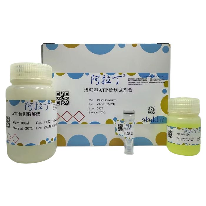
ATP是生物体内能量转换最基本的载体,其含量的变化直接关系到各器官的能量代谢。ATP作为最重要的能量分子在细胞的各种生理、病理过程中起着重要作用。ATP水平的改变,会影响许多细胞的功能。通常细胞在凋亡、坏死或处于一些毒性状态下,ATP水平会下降,而高葡萄糖刺激等对于一些细胞可以上调细胞内ATP水平。通常ATP水平的下降表明线粒体的功能受损或下降,在细胞凋亡时ATP水平的下降通常和线粒体的膜电位下降同时发生。ATP含量测定试剂盒可以用于检测普通溶液、细胞或组织内的ATP水平。
本试剂盒根据萤火虫荧光素酶(firefly luciferase,也称荧光素酶)催化荧光素产生荧光时需要ATP提供能量研制而成。当萤火虫荧光素酶和荧光素都过量时,在一定的浓度范围内荧光的产生和ATP的浓度成正比。这样就可以高灵敏地检测溶液中的ATP浓度。
E1501756
| Component
| 200T | Storage
|
E1501756A
| ATP检测试剂 | 25 mL
| -20℃. Store in the dark.
|
E1501756B
| ATP标准溶液 | 100 μL | -20℃. Store in the dark.
|
E1501756C
| ATP检测裂解液 | 100 mL | -20℃. Store in the dark. |
产品优势
1、灵敏度高:在0.1nM至100μM范围内有良好的检测结果。
2、稳定性高:30分钟内样品ATP测定结果下降不超过10%。
3、制备的样品兼容性好:使用本试剂盒中的ATP检测裂解液裂解获得的细胞或组织样品,不仅可以用于ATP检测,还可用于检测蛋白浓度、进行SDS-PAGE或一些常规的较易溶解蛋白的Western检测。
4、使用方便快捷:通常10-20个样品可以在30-60分钟内测定完毕。
5、样品制备简单:样品无需进行高氯酸或三氯乙酸(TCA)抽提,本试剂盒提供了专用裂解液,样品简单裂解后即可用于ATP检测。
实验流程
1、样本制备
注意:样品裂解需在4℃或冰上操作。
1.1 对于贴壁细胞
吸除培养液,按照6孔板每孔加入200μL裂解液的比例(即相当于细胞培养液量2mL的1/10)加入裂解液,裂解细胞。裂解细胞时为了裂解充分,可以使用移液器进行反复吹打或晃动培养板使裂解液充分接触并裂解细胞。通常细胞在接触裂解液后会立即裂解。裂解后4℃ 10000rpm离心5min,取上清,用于后续的测定。
1.2 对于悬浮细胞
用离心管离心沉淀细胞,弃上清,轻轻弹散细胞,按照6孔板每孔的细胞量加入200μL裂解液的比例加入裂解液,裂解细胞。裂解细胞时为了裂解充分可以弹击离心管管底或适当 Vortex 使裂解液充分接触并裂解细胞。通常细胞在接触裂解液后会立即裂解。裂解后4℃ 10000rpm离心5min,取上清,用于后续的测定。
1.3 对于组织样品
按照每20mg组织加入约100-200μL裂解液的比例加入裂解液,然后用玻璃匀浆器或其它匀浆设备进行匀浆。充分匀浆可以确保组织被完全裂解。裂解后4℃ 10000rpm离心5min,取上清,用于后续的测定。
2、标准曲线制备
冰浴上融解待用试剂,把ATP标准溶液用ATP检测裂解液稀释成适当的浓度梯度。具体的浓度需根据样品中ATP的浓度而定。初次检测可以检测0.01、0.03、0.1、0.3、1、3和10μM这几个浓度,在后续的实验中,可以根据样品中ATP的浓度对标准品的浓度范围进行适当调整。
| 编号 | 裂解液体积(µL)
| ATP标准溶液体积
| 最终浓度(μM)
|
| A | 98
| 从ATP标准溶液(0.5mM)中取2µL
| 10 |
| B | 70
| 从A管取30µL
| 3 |
| C | 90
| 从A管取10µL
| 1 |
| D | 90
| 从B管取10µL
| 0.3 |
| E | 90
| 从C管取10µL
| 0.1 |
| F | 90
| 从D管取10µL
| 0.03 |
| G | 90
| 从E管取10µL
| 0.01 |
3、ATP浓度的测定
3.1 加100μL ATP检测试剂到检测孔内,室温放置3-5min。
3.2 在检测孔内加上10μL样品或已稀释好的ATP标准溶液。
3.3 用化学发光仪测定RLU值。
注:样品的体积可以自行在10-100μL范围内调节。如果样品中的ATP浓度比较低则可以加入100 μL样品,如果样品中ATP浓度比较高则可以加入较小体积的样品,同时标准品也需要使用相同的 体积。如果样品中ATP的浓度特别高,可以用ATP检测裂解液稀释样品后再测定。
注意事项
1、本试剂盒的检测试剂中含有荧光素酶,反复冻融会导致其逐渐失活。为取得良好的使用效果,第一次解冻后可适当分装保存,但需注意分装的容器不能有ATP污染。
2、荧光素酶的活性对温度比较敏感,所以反应前细胞和ATP检测试剂均需平衡至室温后再进行测 定。请勿室温存放。
3、ATP,特别是裂解后样品中的ATP在室温不太稳定,需在4℃或冰上操作。
4、检测时需使用适合于细胞培养的白色或黑色的96孔板或384孔板。如果使用普通透明的96孔板 或384孔板,相邻孔之间会产生相互干扰。
5、本试剂盒提供的ATP检测裂解液可以有效裂解并释放常见的培养细胞和组织中的ATP。对于一些特殊的组织或样品,如果发现检测出来的ATP水平显著低于预期水平,可以在裂解样品后并且在离心前,取部分样品煮沸2分钟以充分释放ATP。煮沸后样品中的蛋白会变性,从而会在后续的离心步骤中被沉淀,因此煮沸的样品不能用于蛋白浓度测定、SDS-PAGE和Western检测。可以使用剩余的部分样品进行蛋白浓度测定、SDS-PAGE和Western检测。
6、为了您的安全和健康,操作时请穿着实验服并佩戴一次性手套。
实验数据
1、细胞ATP含量测定结果

2、ATP溶液及Vero细胞中ATP含量测定结果
表1:我司和对照产品对ATP溶液和Vero细胞中ATP含量的测定结果
产品厂家
| ATP溶液(μM)
| Vero细胞(μM)
|
| 我司产品 | 1.53 | 20.27 |
对照产品
| 1.47 | 12.16 |
3、样品稳定性检测

4、本产品在0.01μM-10μM的浓度范围内有很好的线性关系

5、本产品在0.1nM-100nM的浓度范围内有很好的线性关系

图4:本产品对ATP标准品的检测效果(0.1-100nM)
6、裂解后样品用于WB检测实验(以β-actin作为内参)

图5:我司和对照产品裂解HL-60细胞后,样品进行WB实验
实验结论
1、由图1可以看出,使用我司产品及对照产品裂解不同细胞后检测其ATP含量,我司产品检测数值基本优于对照品。
2、由表1可以看出,我司产品和对照产品检测同样浓度的ATP溶液时,结果相近;但检测同样数量细胞时,我司产品测定结果更高,数据更加准确。
3、由图2可以看出,使用我司产品及对照产品持续检测30min样品中ATP含量,我司产品稳定性优于对照品。
4、由图3、图4可以看出,我司产品在加入10μl-100μl标准品进行检测时,在0.1nM-10μM的ATP浓度范围内具有良好的线性关系。
5、由图5可以看出,使用我司产品及对照产品裂解HL-60细胞后样品进行WB实验,我司产品裂解效果优于对照产品。
ATP is the most fundamental energy currency in living organisms, and its concentration directly affects the energy metabolism of various organs. As the most important energy molecule, ATP plays a critical role in diverse physiological and pathological processes. Changes in ATP levels can impact numerous cellular functions. Typically, ATP levels decrease during apoptosis, necrosis, or under certain toxic conditions, while high glucose stimulation can upregulate intracellular ATP levels in some cells. A decrease in ATP levels often indicates impaired or declining mitochondrial function. During apoptosis, the drop in ATP levels usually occurs simultaneously with a decrease in mitochondrial membrane potential. The ATP Assay Kit can be used to detect ATP levels in common solutions, cells, or tissues.
This kit is developed based on the principle that firefly luciferase requires ATP to provide energy for catalyzing the production of light from luciferin. When both firefly luciferase and luciferin are in excess, the light produced is proportional to the ATP concentration within a certain range. This allows for highly sensitive detection of ATP concentration in solutions.
E1501756
| Component
| 200T | Storage
|
E1501756A
| ATP Detection Reagent
| 25 mL
| -20℃. Store in the dark.
|
E1501756B
| ATP Standard Solution | 100 μL | -20℃. Store in the dark.
|
E1501756C
| ATP Assay Lysis Buffer | 100 mL | -20℃. Store in the dark. |
Product Advantages
1. High Sensitivity: Provides excellent detection results within the range of 0.1 nM to 100 μM.
2. High Stability: ATP measurement results from prepared samples decrease by no more than 10% within 30 minutes.
3. Good Compatibility of Prepared Samples: Cell or tissue samples lysed using the ATP Assay Lysis Buffer provided in this kit can not only be used for ATP detection but also for protein concentration assays, SDS-PAGE, or Western blotting for some commonly soluble proteins.
4. Convenient and Fast: Typically, 10-20 samples can be assayed within 30-60 minutes.
5. Simple Sample Preparation: Samples do not require perchloric acid or trichloroacetic acid (TCA) extraction. The specialized lysis buffer provided allows samples to be used for ATP detection after simple lysis.
Experimental Procedure
1. Sample Preparation
Note: Sample lysis should be performed at 4°C or on ice.
1.1 For Adherent Cells
Remove the culture medium. Add Lysis Buffer according to the proportion of 200 μL per well of a 6-well plate (i.e., 1/10 of the 2 mL culture medium volume) to lyse the cells. For complete lysis, pipette up and down repeatedly or shake the plate to ensure the lysis buffer fully contacts and lyses the cells. Cells typically lyse immediately upon contact with the buffer. Centrifuge the lysate at 10,000 rpm, 4°C for 5 minutes. Collect the supernatant for subsequent assay.
1.2 For Suspension Cells
Centrifuge to pellet the cells, discard the supernatant, and gently resuspend the pellet. Add Lysis Buffer according to the proportion of 200 μL per the cell amount from one well of a 6-well plate. For complete lysis, tap the tube bottom or vortex appropriately to ensure the lysis buffer fully contacts and lyses the cells. Cells typically lyse immediately. Centrifuge the lysate at 10,000 rpm, 4°C for 5 minutes. Collect the supernatant for subsequent assay.
1.3 For Tissue Samples
Add Lysis Buffer in a ratio of approximately 100-200 μL per 20 mg of tissue. Homogenize using a glass homogenizer or other homogenization equipment. Thorough homogenization ensures complete tissue lysis. Centrifuge the lysate at 10,000 rpm, 4°C for 5 minutes. Collect the supernatant for subsequent assay.
2. Standard Curve Preparation
Thaw the required reagents on ice. Dilute the ATP Standard Solution with ATP Assay Lysis Buffer to create appropriate concentration gradients. The specific concentrations should be determined based on the expected ATP concentration in the samples. For initial detection, concentrations of 0.01, 0.03, 0.1, 0.3, 1, 3, and 10 μM can be tested. In subsequent experiments, adjust the standard concentration range appropriately based on sample ATP levels.
| Tube | Lysis Buffer Volume (μL)
| ATP Standard Solution Volume
| Final Concentration (μM)
|
| A | 98
| 2 μL from stock (0.5 mM)
| 10 |
| B | 70
| 30 μL from Tube A
| 3 |
| C | 90
| 10 μL from Tube A
| 1 |
| D | 90
| 10 μL from Tube B
| 0.3 |
| E | 90
| 10 μL from Tube C
| 0.1 |
| F | 90
| 10 μL from Tube D
| 0.03 |
| G | 90
| 10 μL from Tube E
| 0.01 |
3. ATP Concentration Measurement
3.1 Add 100 μL of ATP Detection Reagent to each assay well. Incubate at room temperature for 3-5 minutes.
3.2 Add 10 μL of sample or the diluted ATP standard solution to the assay well.
3.3 Measure the Relative Light Unit (RLU) value using a luminometer.
Note: The sample volume can be adjusted within the range of 10-100 μL. If the ATP concentration in the sample is low, 100 μL can be added. If the ATP concentration is high, a smaller volume can be used, but the same volume must be used for the standard curve samples. If the ATP concentration is exceptionally high, dilute the sample with ATP Assay Lysis Buffer before measurement.
Precautions
1. The Detection Reagent contains luciferase. Repeated freeze-thaw cycles will lead to gradual inactivation. For optimal performance, consider aliquoting after the first thaw, ensuring the aliquot containers are free from ATP contamination.
2. Luciferase activity is temperature-sensitive. Before the reaction, equilibrate both cells and the ATP Detection Reagent to room temperature for measurement. Do not store at room temperature for extended periods.
3. ATP, especially in lysed samples, is unstable at room temperature. Perform operations at 4°C or on ice.
4. Use white or black 96-well or 384-well plates suitable for cell culture for detection. Using standard transparent plates may cause interference between adjacent wells.
5. The provided ATP Assay Lysis Buffer effectively lyses and releases ATP from common cultured cells and tissues. For special tissues or samples where detected ATP levels are significantly lower than expected, boil a portion of the lysate for 2 minutes before centrifugation to fully release ATP. Boiling will denature proteins, which will precipitate during subsequent centrifugation; therefore, boiled samples cannot be used for protein concentration assays, SDS-PAGE, or Western blotting. Use the remaining portion of the sample for protein assays, SDS-PAGE, and Western blotting.
6. For your safety and health, wear a lab coat and disposable gloves during operation.

 ZJ25F1029228


 危险品化学品经营许可证(带存储)